Transcription of Clinical Comparison of Sclerotherapy Versus Long …
1 Clinical Comparison of Sclerotherapy Versus long -Pulsed Nd:YAG Laser Treatment for Lower Extremity Telangiectases Jason R. Lupton, MD, Tina S. Alster, MD, and Patti Romero, RN, BSN. Washington Institute of Dermatologic Laser Surgery, Washington, background. Sclerotherapy has traditionally been considered Nd:YAG laser irradiation on the other. Patients were evaluated the gold standard of treatment for leg veins, but patient fear of by two masked assessors at each treatment visit and at 1 and 3. multiple needle injections and side effects of treatment have fu- months after treatment to assess Clinical improvement within eled investigation into other treatment alternatives. As a result, matched sites. vascular-specific laser and light sources have been developed in results. Leg telangiectases responded best to Sclerotherapy in an effort to treat these vessels with minimal morbidity and im- fewer treatment sessions than to long -pulsed 1064 nm Nd:YAG.
2 Proved efficacy. laser irradiation. The incidence of adverse sequelae was mini- objective. To compare the Clinical efficacy of leg telangiecta- mal and equivocal in both treatment groups. sia treatment with sodium tetradecyl sulfate Sclerotherapy to conclusion. Despite recent advances in laser technology for long -pulsed 1064 nm Nd:YAG laser irradiation. treatment of lower extremity telangiectases, Sclerotherapy con- methods. A series of 20 patients with size-matched superficial tinues to offer superior Clinical effect in the majority of cases. telangiectases of the lower extremities were randomly assigned Laser leg vein treatment appears to be most beneficial in pa- to receive two consecutive monthly treatments with injectable tients with telangiectatic matting, needle phobia, or sclerosant sodium tetradecyl sulfate on one leg and long -pulsed 1064 nm allergy.
3 J. R. LUPTON, MD, T. S. ALSTER, MD, AND P. ROMERO, RN, BSN HAVE INDICATED NO SIGNIFICANT INTEREST. WITH COMMERCIAL SUPPORTERS. APPROXIMATELY 40% of women and 15% of men tense pulsed light (IPL) sources have recently been develop clinically apparent ectatic venules of the lower developed and have shown early promise in the treat- legs,1 with more than half of them becoming symp- ment of leg veins up to 3 mm in 11. tomatic over Because leg veins are located rela- With its relatively long wavelength of 1064 nm and tively deep in the dermis, with increased hydrostatic subsequent decreased absorption of energy by epider- pressures, large luminal diameters with thick vessel mal melanin, the long -pulsed Nd:YAG laser may best walls, as well as decreased blood oxygenation, they be able to penetrate tissue to a level deep enough to ef- are more difficult to eradicate than superficial facial fect destruction of leg ,12,13 This study vessels.
4 The vascular anatomy of the legs is also was conducted to compare the effectiveness of sclero- unique in that superficial vessels are interconnected therapy Versus long -pulsed 1064 nm Nd:YAG laser ir- with deeper, reticular veins and thus usually require radiation of lower extremity telangiectases. more extensive treatment even if obvious varicosities are not ,4. Materials and Methods Although Sclerotherapy is considered the gold stan- dard of leg vein treatment by most practitioners, it is Twenty women, ages 27 68 years (mean 45 years), skin associated with several limitations, including the pos- phototypes I III, with size-matched superficial leg telang- sibility of systemic allergic reactions to the injected iectases (diameter range mm; mean mm) were sclerosant, posttreatment ulceration, scarring, telang- included for study. Patients with a prior history of lower ex- iectatic matting, and the need for multiple transcuta- tremity telangiectasia treatment, Clinical evidence of severe neous ,6 A variety of different laser and in- vascular incompetence, on anticoagulant treatment, or those currently pregnant or breastfeeding were excluded from the study.
5 Address correspondence and reprint requests to: Tina S. Alster, MD, Patients were randomized to receive two treatments with 2311 M St. NW, Suite 200, Washington, 20037, or e-mail: talster@ a long -pulsed 1064 nm Nd:YAG laser (Varia, CoolTouch Laser Corp., Auburn, LA) to telangiectases on one leg and 2002 by the American Society for Dermatologic Surgery, Inc. Published by Blackwell Publishing, Inc. ISSN: 1076-0512/02/$ Dermatol Surg 2002;28:694 697. Dermatol Surg 28:8:August 2002 lupton et al.: sclerotherpay Versus nd:yag laser 695. sodium tetradecyl sulfate (Sotradecol, Elkins-Sinn Inc., Cherry Hill, NJ) Sclerotherapy to those on the other. Size-matched vessels on the thighs, knees, calves, ankles, and popliteal fossae received treatment by a single operator ( ). Laser treatments were delivered through a mm collimated spot size at 1 Hz using fluences of 125 150 J/cm2.
6 (mean 135 J/cm2). A pulse duration of 25 msec was used for smaller vessels and a 50-msec pulse width was applied for vessels larger than mm in diameter. Epidermal cooling was achieved with a cryogen spray of varying pre- and post- treatment durations depending upon the skin phototype of the patient (ie, longer precooling with darker skin photo- types) and the size of the vessel (ie, increased postcooling de- lay for larger vessels in order to effect full-thickness mural denaturation). Precooling durations ranged from 0 to 5. msec, postcooling durations ranged from 20 to 50 msec, and postcooling delays ranged from 5 to 20 msec. The end point of laser treatment was determined to be when either total contraction (visual elimination) of the vessel was effected or darkening of the vessel and/or adjacent tissue hyperemia was observed. Size-matched vessels on the contralateral leg were in- jected with sodium tetradecyl sulfate through a 30- gauge needle.
7 A sufficient amount of injectable sclerosant was delivered to completely blanch the targeted vessels and all visible tributaries during the treatment session (range 2 10 ml). Graduated compression stockings of 20 30. mmHg were applied on the Sclerotherapy -treated leg for 48. hours following treatment. Photographic documentation and Clinical improvement scores were determined 1 month after the first treatment ses- sion, and 1 and 3 months after the second treatment session by two masked independent assessors using a quartile grad- ing scale of 0 less than 25% improvement, 1 26 50%. improvement, 2 51 75% improvement, and 3 greater than 75% improvement. Side effects of treatment were also recorded at each treatment and follow-up visit. Results Clinical Improvement Average Clinical improvement scores were for the Sclerotherapy -treated leg and for the laser-treated leg 1 month after the first treatment session (Figures 1 3).
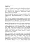
8 One month following the second treatment ses- sion, improvement scores of and were given for the Sclerotherapy -treated side and the laser-treated side, respectively. Three months after the second treat- ment session, average improvement scores of and were given for the Sclerotherapy -treated side and laser-treated side, respectively. Figure 1. A) Medial malleolus with telangiectasias measuring . mm in diameter. B) One month after the first Sclerotherapy ses- Side Effects sion with sodium tetradecyl sulfate solution. C) Three months after the second treatment with sodium tetradecyl sulfate. In general, both the laser treatments and the sclero- therapy were well tolerated by all patients, with no se- 696 lupton et al.: sclerotherpay Versus nd:yag laser Dermatol Surg 28:8:August 2002. Figure 3. Clinical improvement scores. rious adverse sequelae.
9 Of the 20 patients treated, 70% (n 14) reported mild treatment pain associated with both treatments. Transient local tissue erythema and edema lasting between 1 and 7 days were re- ported by 95% of patients (n 19) on the sclerother- apy-treated side and by 75% of patients (n 15) on the laser-treated side. Transient postinflammatory hy- perpigmentation developed in two patients (10%) on the Sclerotherapy -treated leg, both of whom had skin phototype III, but in no patients who received laser treatment. There were no cases of vesiculation, fibro- sis, or scarring as a result of either treatment. Discussion Significant Clinical improvement of leg telangiectases was shown in all sites treated with either Sclerotherapy or laser irradiation. Earlier vessel clearing was ob- served after Sclerotherapy , with higher average im- provement scores in Sclerotherapy -treated areas at each follow-up visit.
10 Posttreatment hyperpigmentation observed on the Sclerotherapy side may have been a result of the relatively high concentration ( ) of sclerosant used and potentially could have been avoided with injection of a less concentrated solution ( ). Given the fact that laser treatment of lower extrem- ity telangiectases is not associated with either im- proved Clinical efficacy or decreased treatment pain compared with traditional Sclerotherapy , its wide- spread use cannot yet be supported for this purpose. Although Sclerotherapy is relatively inexpensive, it re- quires extensive Clinical experience to achieve optimal results. The novice practitioner may find laser treat- ment easier to perform and may therefore initially pre- fer this method of treatment. Figure 2. A) Contralateral ankle of the patient from Figure 2, dem- onstrating fine mm diameter telangiectasias.