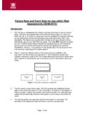
Transcription of Clinical corner Revisiting respiratory failure - www.hcpro.com
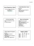
1 20 January 2014 2014 hcpro , a division of permission to reproduce part or all of this newsletter for external distribution or use in educational packets, please contact the Copyright Clearance Center at or cornerRevisiting respiratory failure by Richard D. Pinson, MD, FACP, CCSThe diagnosis and documentation of respiratory failure continues to be challeng-ing for coders, documentation specialists, and physicians. Many physicians, including pulmon-ologists, are unaware of the current Clinical standards for diag-nosing acute respiratory failure and commonly overlook the presence of chronic respiratory failure . Yet they typically iden-tify multiple Clinical criteria and provide appropriate manage-ment for respiratory failure , which creates query opportunities. In this article, we will discuss a variety of Clinical indica-tors for respiratory failure and identify a number of com-mon documentation improvement of acute respiratory failureAcute respiratory failure is classified as hypoxemic (low arterial oxygen levels), hypercapneic (elevated levels of car-bon dioxide gas), or a combination of the two.
2 In most cases one or the other predominates. For ICD-9, these terms, being nonessential modifiers, are irrelevant for code assign-ment. ICD-10, however, has codes that permit a distinction (see Table 1), but the distinction is not a requirement and queries for it will not alter its MCC classification. The clini-cal criteria for diagnosing acute respiratory failure are: Hypoxemic: Partial pressure of oxygen (pO2) level less than (<) 60 millimeter(s) of mercury (mmHg) (oxygen saturation [SpO2] < 91%) on room air, or pO2/frac-tion of inspired oxygen (FIO2) (P/F) ratio < 300, or 10 mmHg decrease in baseline pO2 (if known) Hypercapnic: Partial pressure of carbon dioxide (pCO2) >50 mmHg with pH < , or 10 mmHg increase in baseline pCO2 (if known)With the exception of the P/F ratio, these criteria have also been offered as assistance to coders and documentation specialists for recognizing possible acute respiratory failure (see AHA s Coding Clinic for ICD-9-CM, Third Quarter 1988, p.)
3 7; and Second Quarter 1990, p. 20).Management that requires endotracheal intubation and mechanical ventilation or initiation of biphasic positive air-way pressure (BiPAP) nearly always means the patient has acute respiratory failure , but these measures are not required for the diagnosis. Similarly, providing 40% or more supple-mental oxygen implies that the physician is treating acute respiratory failure since only a patient with acute respiratory failure would need that much hypoxemic respiratory failureThe gold standard for the diagnosis of hypoxemic respiratory failure is an arterial pO2 on room air less than 60 mmHg measured by arterial blood gases (ABG). In the absence of an ABG, SpO2 measured by pulse oximetry on room air can serve as a substitute for the pO2: SpO2 of 91% equals pO2 of 60 mmHg. These criteria may not apply to patients with chronic respiratory failure ( , severe chron-ic obstructive pulmonary disease [COPD]), because their room air pO2 is often less than 60 mmHg (SpO2 < 91%).
4 Chronic respiratory failure patients are treated with supplemental oxygen on a continuous outpatient basis to keep arterial oxygen above these levels. However, if the baseline pO2 is known, a decrease by 10 mmHg or more indicates acute hypoxemic respiratory failure in such a P/F ratioThe P/F ratio is a powerful objective tool to identify acute hypoxemic respiratory failure at any time while the patient is receiving supplemental oxygen, a frequent problem faced by documentation specialists where no room air ABG is available, or pulse oximetry readings seem equivocal. The P/F ratio equals the arterial pO2 ( P ) from the ABG divided by the FIO2 ( F ) the fraction (percent) of inspired oxygen that the patient receives expressed as a decimal (40% oxygen = FIO2 of ). A P/F ratio less than 300 indicates acute respiratory failure .
5 Most physicians have never heard of the P/F ratio, but it was validated and has been used in the context of acute respiratory distress syndrome (ARDS) for many years, where acute respiratory failure is called acute lung injury. A P/F ratio < 300 indicates mild ARDS, < 200 is consistent with moderate ARDS, and < 100 is severe ARDS. The P/F ratio indicates what the pO2 would be on room air: P/F ratio < 300 = a pO2 < 60 mm Hg on room air 2014 hcpro , a division of BLR. January 2014 21 For permission to reproduce part or all of this newsletter for external distribution or use in educational packets, please contact the Copyright Clearance Center at or 978-750-8400. P/F ratio < 250 = a pO2 < 50 mm Hg on room air P/F ratio < 200 = a pO2 < 40 mm Hg on room air As an example, suppose the pO2 is 90 mmHg on 40% oxygen (FIO2 =.)
6 40). The P/F ratio = 90 divided by .40 = 225 (rather severe acute respiratory failure ). The pO2 on room air in this case would have been about 45 mmHg (well below the cutoff of 60 mmHg).The validity of the P/F ratio is not limited to ARDS. It simply expresses a consistent physiologic relationship between inspired oxygen and arterial pO2 regardless of cause. Authoritative applications of the P/F ratio in set-tings other than ARDS include pneumonia and sepsis. The Infectious Disease Society of America and the American Thoracic Society recognize a P/F ratio less than 250 as one of the 10 criteria for severe community-acquired pneumonia that may require admission to intensive care. The International Sepsis Definition criteria (2001) and the Surviving Sepsis - Severe Sepsis Guidelines (2008 and 2012) use a P/F ratio < 300 as an indicator of acute organ ( respiratory ) may be translated to pO2 The arterial pO2 measured by ABG is the definitive method for calculating the P/F ratio.
7 However, when the pO2 is unknown because an ABG is not available, the SpO2 measured by pulse oximetry can be used to approximate the pO2, as shown in Table 2. It is important to note that esti-mating the pO2 from the SpO2 becomes unreliable when the SpO2 is greater than 97%. For example, suppose a patient on 40% oxygen has a pulse oximetry SpO2 of 95%. Referring to Table 2, SpO2 of 95% is equal to a pO2 of 80 mmHg. The P/F ratio = 80 divided by = 200 (quite severe acute respiratory failure ). The patient may be stable receiving 40% oxygen, but still has acute respiratory failure . If oxygen were withdrawn, leaving her on room air, the pO2 would only be 42 mmHg (much less than 60 mmHg on room air).Translating supplemental oxygen Supplemental oxygen may be administered by mask or nasal cannula. A Venturi mask (Venti-mask) delivers a controlled flow of oxygen at a specific fixed concentration (FIO2): 24%, 28%, 31%, 35%, 40%, and 50%.
8 The non-rebreather (NRB) mask is designed to deliver approximately 100% oxygen. Providing 40% or more supplemental oxygen implies that the physician is treating acute respiratory failure since only such a patient would need that much oxygen. A nasal cannula provides oxygen at adjustable flow rates in liters of oxygen per minute (L/min or LPM). The actual FIO2 (percent oxygen) delivered by nasal cannula is some-what variable and less reliable than with a mask, but can be estimated as shown in Table 3. The FIO2 derived from nasal cannula flow rates can then be used to calculate the P/F ratio. For example, a patient has a pO2 of 85 mmHg on ABG while receiving 5 L/min of oxygen. Since 5 L/min is equal to 40% oxygen (an FIO2 of ), the P/F ratio = 85 divided by = (clearly severe acute respiratory failure ).Acute hypercapneic respiratory failureThe hallmark of acute hypercapneic respiratory failure is Table 1: ICD-10-CM codes for respiratory failureThe following codes are applicable for respiratory failure under ICD-10-CM: : Acute respiratory failure (MCCs) : unspecified whether with hypoxia or hypercapnia : with hypoxia : with hypercapnia : Chronic respiratory failure (CCs) : unspecified whether with hypoxia or hypercapnia : with hypoxia : with hypercapnia : Acute and/on chronic respiratory failure (MCCs) : unspecified whether with hypoxia or hypercapnia : with hypoxia : with hypercapnia : respiratory failure , unspecified : respiratory failure , unspecified, (unspecified whether with hypoxia or hypercapnia) : respiratory failure , unspecified with hypoxia.
9 respiratory failure , unspecified with hypercapnia (excludes newborn, postprocedural, ARDS, respiratory arrest, and cardiorespiratory failure )22 January 2014 2014 hcpro , a division of permission to reproduce part or all of this newsletter for external distribution or use in educational packets, please contact the Copyright Clearance Center at or pCO2 due to retention/accumulation of carbon diox-ide gas resulting in an acidic pH less than There are many causes, but severe COPD is the most common. Physicians can establish a diagnosis by viewing a pCO2 greater than 50 mmHg with a pH less than If the pH is greater than , the patient has chronic (not acute) respiratory failure . Physicians often identify this Clinical condition as respira-tory acidosis, which is the same thing as acute hypercapneic respiratory failure .
10 Unfortunately, the code for respiratory aci-dosis is , which is a CC, in contrast to the MCC status of acute respiratory failure hence the need for , if the baseline pCO2 is known, an increase of 10 mmHg or more indicates acute hypercapneic respiratory failure . Finally, an exacerbation of symptoms requiring an increase in chronic supplemental oxygen indicates an acute exacerbation of chronic respiratory failure , which would be classified as acute-on-chronic respiratory failure if properly respiratory failureChronic respiratory failure is very common in patients with severe COPD and other chronic lung diseases such as cystic fibrosis and pulmonary fibrosis. It is characterized by a combination of hypoxemia, elevated pCO2, elevated bicarbonate level, and normal pH ( ). The most important tip-off to chronic respiratory failure is chronic dependence on supplemental oxygen ( home O2 ).