Transcription of Colon and Rectum - College of American Pathologists
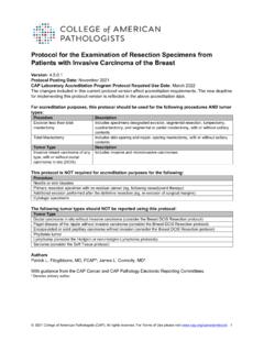
1 Protocol for the Examination of Specimens From Patients With Primary Carcinoma of the Colon and Rectum Version: Colon Rectum Protocol Posting Date: June 2017 Includes pTNM requirements from the 8th Edition, ajcc staging manual For accreditation purposes, this protocol should be used for the following procedures AND tumor types: Procedure Description Colectomy Includes specimens designated total, partial, or segmental resection Rectal Resection Includes specimens designated low anterior resection or abdominoperineal resection Tumor Type Description Carcinoma Invasive carcinomas including small cell and large cell (poorly differentiated) neuroendocrine carcinoma This protocol is NOT required for accreditation purposes for the following: Procedure Excisional biopsy (polypectomy) Local excision (transanal disk excision) Primary resection specimen with no residual cancer (eg, following neoadjuvant therapy) Cytologic specimens The following tumor types should NOT be reported using this protocol: Tumor Type Well-differentiated neuroendocrine tumors (consider the Colorectal NET protocol) Lymphoma (consider the Hodgkin or non-Hodgkin Lymphoma protocol) Sarcoma (consider the Soft Tissue protocol) Authors Sanjay Kakar, MD*; Chanjuan Shi MD, PhD*; Mariana E.
2 Berho, MD, PhD; David K. Driman, MBchB; Patrick Fitzgibbons, MD; Wendy L. Frankel, MD; Kalisha A. Hill, MD, MBA; John Jessup, MD; Alyssa M. Krasinskas, MD; Mary K Washington, MD, PhD With guidance from the CAP Cancer and CAP Pathology Electronic Reporting Committees. * Denotes primary author. All other contributing authors are listed alphabetically. 2017 College of American Pathologists (CAP). All rights reserved. For Terms of Use please visit Gastrointestinal Colon and Rectum ColonRectum Accreditation Requirements This protocol can be utilized for a variety of procedures and tumor types for clinical care purposes. For accreditation purposes, only the definitive primary cancer resection specimen is required to have the core and conditional data elements reported in a synoptic format. Core data elements are required in reports to adequately describe appropriate malignancies. For accreditation purposes, essential data elements must be reported in all instances, even if the response is not applicable or cannot be determined.
3 Conditional data elements are only required to be reported if applicable as delineated in the protocol. For instance, the total number of lymph nodes examined must be reported, but only if nodes are present in the specimen. Optional data elements are identified with + and although not required for CAP accreditation purposes, may be considered for reporting as determined by local practice standards. The use of this protocol is not required for recurrent tumors or for metastatic tumors that are resected at a different time than the primary tumor. Use of this protocol is also not required for pathology reviews performed at a second institution (ie, secondary consultation, second opinion, or review of outside case at second institution). Transanal disk excision is NOT considered to be the definitive resection specimen for the purpose of cancer reporting, even though the entire cancer may be removed. A protocol is recommended for reporting such specimens for clinical care purposes, but this is not required for accreditation purposes.
4 Synoptic Reporting All core and conditionally required data elements outlined on the surgical case summary from this cancer protocol must be displayed in synoptic report format. Synoptic format is defined as: Data element: followed by its answer (response), outline format without the paired "Data element: Response" format is NOT considered synoptic. The data element must be represented in the report as it is listed in the case summary. The response for any data element may be modified from those listed in the case summary, including Cannot be determined if appropriate. Each diagnostic parameter pair (Data element: Response) is listed on a separate line or in a tabular format to achieve visual separation. The following exceptions are allowed to be listed on one line: o Anatomic site or specimen, laterality, and procedure o Pathologic Stage Classification (pTNM) elements o Negative margins, as long as all negative margins are specifically enumerated where applicable The synoptic portion of the report can appear in the diagnosis section of the pathology report, at the end of the report or in a separate section, but all Data element: Responses must be listed together in one location Organizations and Pathologists may choose to list the required elements in any order, use additional methods in order to enhance or achieve visual separation, or add optional items within the synoptic report.
5 The report may have required elements in a summary format elsewhere in the report IN ADDITION TO but not as replacement for the synoptic report all required elements must be in the synoptic portion of the report in the format defined above. CAP Laboratory Accreditation Program Protocol Required Use Date: March 2018* * Beginning January 1, 2018, the 8th edition ajcc staging manual should be used for reporting pTNM. CAP Colon and Rectum Protocol Summary of Changes The following data elements were modified: Pathologic Stage Classification (pTNM, AJCC 8th Edition) Histologic Type Histologic Grade Type of Polyp in Which Invasive Carcinoma Arose Additional Pathological Findings The following data element was added: Tumor budding 2 Gastrointestinal Colon and Rectum ColonRectum Resection, Including Transanal Disk Excision of Rectal Neoplasms The following data elements were modified: Histologic Type Histologic Grade Tumor Extension Margins Pathologic Stage Classification (pTNM, AJCC 8th Edition) Type of Polyp in Which Invasive Carcinoma Arose Additional Pathologic Findings The following data element was added: Peritumoral tumor budding The following data element was removed: Histologic Features Suggestive of Microsatellite Instability 3 CAP Approved Gastrointestinal Colon and Rectum ColonRectum Surgical Pathology Cancer Case Summary Protocol posting date.
6 June 2017 Colon AND Rectum : Excisional Biopsy (Polypectomy) Note: This case summary is recommended for reporting biopsy specimens, but is not required for accreditation purposes. Select a single response unless otherwise indicated. Tumor Site (Note A) ___ Cecum ___ Ileocecal valve ___ Right (ascending) Colon ___ Hepatic flexure ___ Transverse Colon ___ Splenic flexure ___ Left (descending) Colon ___ Sigmoid Colon ___ Rectosigmoid region ___ Rectum ___ Other (specify): _____ ___ Not specified + Specimen Integrity + ___ Intact + ___ Fragmented + Polyp Size + Greatest dimension (centimeters): ___ cm + Additional dimensions (centimeters): ___ x ___ cm + ___ Cannot be determined (explain): _____ + Polyp Configuration + ___ Pedunculated with stalk + Stalk length (centimeters): ___ cm + ___ Sessile + Size of Invasive Carcinoma + Greatest dimension (centimeters): ___ cm + Additional dimensions (centimeters): ___x ___ cm + ___ Cannot be determined (explain): _____ Histologic Type (select all that apply) (Note B) ___ Adenocarcinoma ___ Mucinous adenocarcinoma ___ Signet-ring cell carcinoma ___ Medullary carcinoma ___ Micropapillary carcinoma ___ Serrated adenocarcinoma ___ Large cell neuroendocrine carcinoma ___ Small cell neuroendocrine carcinoma ___ Neuroendocrine carcinoma (poorly differentiated)# ___ Squamous cell carcinoma + Data elements preceded by this symbol are not required for accreditation purposes.
7 These optional elements may be clinically important but are not yet validated or regularly used in patient management. 4 CAP Approved Gastrointestinal Colon and Rectum ColonRectum ___ Adenosquamous carcinoma ___ Spindle cell carcinoma ___ Mixed adenoneuroendocrine carcinoma ___ Undifferentiated carcinoma ___ Other histologic type not listed (specify): _____ ___ Carcinoma, type cannot be determined # Note: Select this option only if large cell or small cell cannot be determined Histologic Grade (Note C) ___ G1: Well differentiated ___ G2: Moderately differentiated ___ G3: Poorly differentiated ___ G4: Undifferentiated ___ Other (specify): _____ ___ GX: Cannot be assessed ___ Not applicable Tumor Extension (Note D) ___ Tumor invades lamina propria ___ Tumor invades muscularis mucosae ___ Tumor invades submucosa ___ Tumor invades muscularis propria ___ Cannot be assessed Margins (select all that apply) Deep Margin (Stalk Margin) ___ Cannot be assessed ___ Uninvolved by invasive carcinoma Distance of invasive carcinoma from margin (millimeters or centimeters).
8 ___ mm or ___ cm ___ Involved by invasive carcinoma Mucosal Margin (required only if applicable) ___ Cannot be assessed ___ Uninvolved by invasive carcinoma ___ Involved by invasive carcinoma ___ Involved by adenoma Lymphovascular Invasion (Notes D and E) ___ Not identified ___ Present + ___ Small vessel lymphovascular invasion + ___ Large vessel (venous) invasion ___ Cannot be determined + Tumor Budding (Note F) + ___ Number of tumor buds in 1 hotspot field (e specify total number in area= mm2): _____ + ___ Low score (0-4) + ___ Intermediate score (5-9) + ___ High score (10 or more) + ___ Cannot be determined + Data elements preceded by this symbol are not required for accreditation purposes. These optional elements may be clinically important but are not yet validated or regularly used in patient management. 5 CAP Approved Gastrointestinal Colon and Rectum ColonRectum + Type of Polyp in Which Invasive Carcinoma Arose (Note G) + ___ Tubular adenoma + ___ Villous adenoma + ___ Tubulovillous adenoma + ___ Traditional serrated adenoma + ___ Sessile serrated adenoma/sessile serrated polyp + ___ Hamartomatous polyp + ___ Other (specify): _____ + Additional Pathologic Findings (select all that apply) + ___ None identified + ___ Ulcerative colitis + ___ Crohn disease + ___ Other polyps (type[s]): _____ + ___ Other (specify): _____ + Ancillary Studies (Note N) Note: For reporting molecular testing and immunohistochemistry for mismatch repair proteins, and for other cancer biomarker testing results, the CAP Colorectal Biomarker Template should be used.
9 Pending biomarker studies should be listed in the Comments section of this report. + Comment(s) + Data elements preceded by this symbol are not required for accreditation purposes. These optional elements may be clinically important but are not yet validated or regularly used in patient management. 6 CAP Approved Gastrointestinal Colon and Rectum ColonRectum Surgical Pathology Cancer Case Summary Protocol posting date: June 2017 Colon AND Rectum : Resection, Including Transanal Disk Excision of Rectal Neoplasms Note: This case summary is recommended for reporting transanal disc excision specimens, but is not required for accreditation purposes. Select a single response unless otherwise indicated. Procedure ___ Right hemicolectomy ___ Transverse colectomy ___ Left hemicolectomy ___ Sigmoidectomy ___ Low anterior resection ___ Total abdominal colectomy ___ Abdominoperineal resection ___ Transanal disk excision (local excision) ___ Endoscopic mucosal resection ___ Other (specify): _____ ___ Not specified Tumor Site (select all that apply) (Note A) ___ Cecum ___ Ileocecal valve ___ Right (ascending) Colon ___ Hepatic flexure ___ Transverse Colon ___ Splenic flexure ___ Left (descending) Colon ___ Sigmoid Colon ___ Rectosigmoid region ___ Rectum ___ Colon , not otherwise specified ___ Cannot be determined (explain): _____ + Tumor Location (applicable only to rectal primaries) (Note A) + ___ Entirely above the anterior peritoneal reflection + ___ Entirely below the anterior peritoneal reflection + ___ Straddles the anterior peritoneal reflection + ___ Not specified Tumor Size Greatest dimension (centimeters): ___ cm + Additional dimensions (centimeters).
10 ___ x ___ cm ___ Cannot be determined (explain): _____ Macroscopic Tumor Perforation (Note H) ___ Not identified ___ Present ___ Cannot be determined + Data elements preceded by this symbol are not required for accreditation purposes. These optional elements may be clinically important but are not yet validated or regularly used in patient management. 7 CAP Approved Gastrointestinal Colon and Rectum ColonRectum + Macroscopic Intactness of Mesorectum (if applicable) (Note I) + ___ Complete + ___ Near complete + ___ Incomplete + ___ Cannot be determined Histologic Type (Note B) ___ Adenocarcinoma ___ Mucinous adenocarcinoma ___ Signet-ring cell carcinoma ___ Medullary carcinoma ___ Micropapillary carcinoma ___ Serrated adenocarcinoma ___ Large cell neuroendocrine carcinoma ___ Small cell neuroendocrine carcinoma ___ Neuroendocrine carcinoma (poorly differentiated)# ___ Squamous cell carcinoma ___ Adenosquamous carcinoma ___ Undifferentiated carcinoma ___ Other histologic type not listed (specify): _____ ___ Carcinoma, type cannot be determined # Note: Select this option only if large cell or small cell cannot be determined Histologic Grade (Note C) ___ G1: Well differentiated ___ G2: Moderately differentiated ___ G3: Poorly differentiated ___ G4: Undifferentiated ___ Other (specify): _____ ___ GX.


