Transcription of Combined Pulmonary Fibrosis and Emphysema …
1 5. Combined Pulmonary Fibrosis and Emphysema (CPFE). Keisaku Fujimoto1 and Yoshiaki Kitaguchi2. 1 Department of Clinical Laboratory Sciences, Shinshu University School of Health Sciences. 21st Department of Internal Medicine, Shinshu University School of Medicine. Nagano, Japan 1. Introduction Combined Pulmonary Fibrosis and Emphysema (CPFE) is one of smoking-related lung diseases. Emphysema is characterized by the permanent abnormal enlargement of airspaces distal to the terminal bronchioles, accompanied by destruction of their walls. The characteristics of Emphysema do not, by definition, include thickening of the alveolar septa and Fibrosis . However, coincidental idiopathic Pulmonary Fibrosis (IPF) and Emphysema was firstly reported in 1990 by Wiggins et al (Wiggins J, et al.)
2 , 1990) in London. Smoking-related interstitial lung diseases (SRILD) include desquamative interstitial pneumonia (DIP), respiratory bronchiolitis-related interstitial lung disease (RB-ILD), Pulmonary Langerhans' cell histiocytosis (LCH) and idiopathic Pulmonary Fibrosis (IPF) (Ryu JH, et al., 2001). Tobacco smoking is also major course of Emphysema and chronic obstructive Pulmonary disease (COPD). Smoking is a common risk factor for both Emphysema and Pulmonary Fibrosis . Recently, the occurrence of both Emphysema and Pulmonary Fibrosis in the same patient has received increased attention as the syndrome of Combined Pulmonary Fibrosis and Emphysema (CPFE) (Cottin V, et al., 2005).
3 It has been demonstrated that CPFE syndrome is not rare because on a series of 110 patients with IPF, 28% of them with at least 10% of the lung affected with Emphysema , and thus are considered to have CPFE (Mejia M, et al., 2009). 2. Clinical characteristics of CPFE. In Japan Hiwatari et al (Hiwatari N, et al., 1993) reported nine patients with Pulmonary Emphysema and IPF among 152 Pulmonary Emphysema patients in 1993. Those patients were all men and heavy smokers. Odani et al (Odani K, et al., 2004) reported 31 patients Combined with Pulmonary Emphysema and IPF among 14900 patients who underwent chest CT from January 1996 to March 2001 at Kochi Medical School Hospital in Japan.
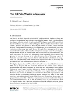
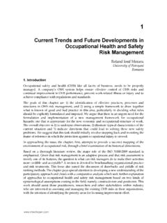
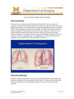
4 The CT of all patients showed the coexistence of Emphysema with upper lung field predominance and diffuse parenchymal lung disease with significant Pulmonary Fibrosis predominantly in the lower lung fields (Figure 1). Centriacinar Emphysema was present in 24 of the 31 (77%). 80 Emphysema patients and paraseptal Emphysema in 11 of 31 patients (35%). Honeycombing, which is one of the most common findings of usual interstitial pneumonia, was present in 24 of the 31. patients (77%). In 2005 Cottin et al (Cottin V, et al., 2005) conducted a retrospective study of 61 patients with CPFE and characterized this association as a distinct entity. They reported that sixty patients were male, dyspnea on exertion was present in all patients, basal crackles were found in 87% and finger clubbing in 43%.
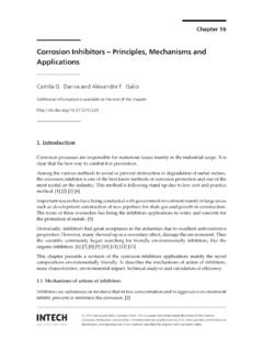
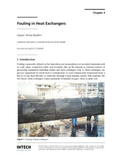
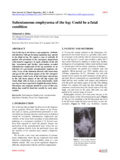
5 Pulmonary function tests showed preserved lung volumes and strongly impaired lung diffusing capacity for carbon monoxide (DLCO). They showed that Pulmonary hypertension was frequent in patients with CPFE with 47% of patients with estimated systolic right ventricular pressure 45 mmHg at echocardiography and the 5-yr probability of survival was 25% in patients with Pulmonary hypertension compared with 75% in those without Pulmonary hypertension at diagnosis (Figure 2). They also confirmed these findings by right heart catheterization on a retrospective multicenter study and concluded that the patients with CPFE and Pulmonary hypertension have a dismal prognosis despite moderately altered lung volume and flows (Cottin V, et al.)
6 , 2010). The risk of developing Pulmonary hypertension is much higher in CPFE than in IPF without Emphysema (Mejia M, et al., 2009). Pulmonary hypertension was a critical determinant of the prognosis. Fig. 1. Imaging in a 66-year-old male with Combined Pulmonary Fibrosis and Emphysema (CPFE). Chest high-resolution computed tomography of the bilateral upper lung fields (a). shows centriaciner + paraseptal Emphysema with thick walled bulla and of the bilateral lower lung fields (b) shows reticular opacities and traction bronchiectasis. Combined Pulmonary Fibrosis and Emphysema (CPFE) 81. Fig. 2. Total survival of patients with Combined Pulmonary Fibrosis and Emphysema (CPFE). (a); 5-yr survival was 55%.
7 Survival for subjects with CPFE stratified on the basis of Pulmonary hypertension (PH) (b); 5-yr survival was significantly less in patients with Pulmonary hypertension (25%) compared with those without Pulmonary hypertension (75%) at diagnosis. We retrospectively examined the clinical characteristics of 47 patients with CPFE based on the findings of chest HRCT consecutively recruited from outpatients attending Shinshu University Hospital between October 2004 and June 2007 (Kitaguchi Y, et al., 2010). The clinical characteristics of CPFE patients were compared with those of Emphysema -dominant COPD patients without parenchymal lung disease (COPD without Fibrosis ). Forty-six of the 47 CPFE patients were male.
8 Paraseptal Emphysema was particularly common in the CPFE. group, although centriacinar was dominant in the COPD without Fibrosis (Table 1). All CPFE patients showed the coexistence of Emphysema with upper lung fields predominance and diffuse parenchymal lung disease with significant Pulmonary Fibrosis with lower lung 82 Emphysema fields predominance. Honeycombing, ground-glass opacities and reticular opacities were present in , and of CPFE patients, respectively. Thick-walled bullae were characteristic in CPFE and observed in more than one half of the CPFE patients. Twenty-seven of the 47 CPFE patients ( ) showed a FEV1/FVC ratio within the normal range and the other patients showed milder airflow limitation, although the presence of severe Emphysema (Table 2, 3).
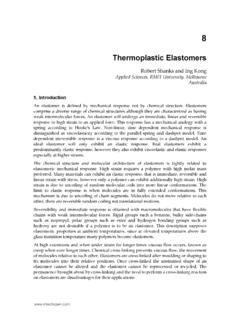
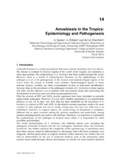
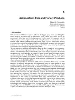
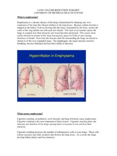
9 All CPFE patients showed lower lung volume and DLCO. than the patients with COPD without Fibrosis as previously reported. Desaturation during 6-min walking test in CPFE patients tended to be more severe than in COPD without Fibrosis patients, if the level of FEV1 or 6-minute walking distance was equal (Figure 3). These findings suggest that CPFE patients show severe dyspnea and severe hypoxemia on effort in spite of subnormal spirometry findings. CPFE COPD. LAA score ** Upper lobe ** Middle lobe ** Lower lobe ** Emphysema type centriacinar, % 11 ( )** 49 ( ). panacinar centriacinar, % 7 ( )* 26 ( ). paraseptal, % 15 ( )** 7 ( ). paraseptal centriacinar, % 12 ( )** 0 ( ). IP distribution Upper lobe 8 ( ).
10 Middle lobe 18 ( ). Lower lobe 47 ( ). IP pattern Thick-walled bulla, n (%) 26 ( ). honeycombing, n (%) 34 ( ). reticular opacity, n (%) 38 ( ). ground glass opacity, n (%) 28 ( ). consolidation, n (%) 6 ( ). traction bronchiectasis, n (%) 18 ( ). peribronchovascular thickening, n (%) 4 ( ). architectural distortion, n (%) 7 ( ). Values are the mean SEM *p< and **p< vs. COPD. Table 1. Chest HRCT findings in patients with Combined Pulmonary Fibrosis and Emphysema (CPFE, n=47) and Emphysema dominant COPD without Fibrosis (COPD, n=82). Combined Pulmonary Fibrosis and Emphysema (CPFE) 83. CPFE COPD. number 47 82. Sex, female/male 1/46 8/74. Body mass index, kg/m2 ** Smoking history, packs year Non-COPD 27 ( ) 0 ( ).