Transcription of Common Causes of False Positive F18 FDG PET/CT …
1 29. , Special Number : pp. 29-35, September 2007. ISSN 1516-8913 Printed in Brazil BRAZILIAN ARCHIVES OF. BIOLOGY AND TECHNOLOGY. A N I N T E R N A T I O N A L J O U R N A L. Common Causes of False Positive F18 FDG PET/CT Scans in Oncology Kevin R. Carter* and Eduard Kotlyarov Michigan State University, POH Medical Center, 50 N. Perry, Pontiac 48342 - Michigan - USA. ABSTRACT. PET/CT is a Common imaging modality used in the evaluation of oncology patients. While being extremely sensitive to identifying sights of malignancy F18 FDG is very non-specific. We attempted to provide a brief review of some of the more Common processes that a nuclear radiology physician may encounter in daily clinical practice that could result in a False Positive diagnosis with F18 FDG PET/CT .
2 A fundamental understanding of the limitations of this technology by the interpreting physician is necessary to avoid making inaccurate diagnosis and potentially limiting important treatments for our patients. Key words: PET/CT , False Positives INTRODUCTION staging of multiple malignancies (Carter and Kotlyarov, 2005). It is approved in the United Radiologic imaging provides an important States for the characterization of solitary component to both the diagnosis and management pulmonary nodules, the diagnosis-staging- of oncology patients. Through the accurate restaging of colon carcinoma, lymphoma, noninvasive visualization of pathologic processes, melanoma, esophageal carcinoma, and head and there is a resulting decrease in the overall neck cancers.
3 (See Fig. 1) It is also useful to morbidity and mortality. Traditionally, evaluate the efficacy of chemotherapy and/or Computerized Tomography (CT) has been utilized radiation therapy following treatment (MNCDM, to evaluate anatomical pathology. This 2007). (See Fig. 2). information was visually combined with the PET/CT offers a unique approach to the diagnosis functional information of Positron Emission and staging of malignancy by exploiting the Tomography (PET) with F18 fluorodioxyglucose biochemical differences between benign and (FDG). Current technology allows for malignant cells Kostakoglu et al., 2003). FDG is simultaneous acquisition of this information which an analog of glucose and is used as a marker of allows precise localization and characterization of glucose metabolism.
4 It is taken up in both tumor pathological processes. Utilization of PET/CT has cells and cells involved in other pathologic been shown to be beneficial in altering the conditions due to an overall increase in the number management of patients compared to imaging with of glucose transporter proteins and increased just CT or PET alone (Mavi et al., 2005). intracellular hexokinase and phosphofructokinase PET/CT is currently utilized in the diagnosis and levels, which promote glycolysis. FDG acts *. Author for correspondence Brazilian Archives of Biology and Technology 30 Carter, K. R. and Kotlyarov, E. similarly to D-glucose in transport through the cell This result in increased uptake of FDG within membrane and phosphorylation by hexokinase.
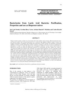
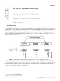
5 Metabolically active cells. Sites of physiologic Once FDG is phosphorylated, hexose-phosphate accumulation of FDG include organs which enzyme prevents FDG from being further normally utilize glucose for metabolism (brain, catabolized or transported back into extracellular muscles striated and smooth, salivary glands, space in substantial amounts, essentially trapping myocardium, gastrointestinal system, urinary the FDG (Kostakoglu et al., 2003). system, thyroid gland, and gonadal issues). B. C. A. Figure 1 - This 56 year old male presented with an abnormal pre-employment screening chest x- ray. The PET/CT was performed to determine if an abnormality present in the lung was malignant or benign.
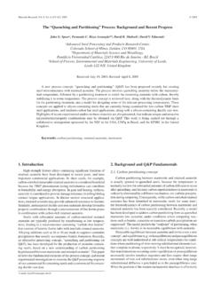
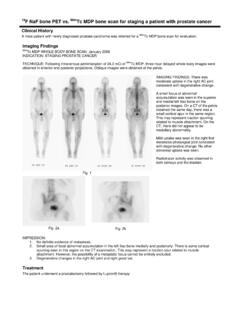
6 Whole body maximum intensity projection (MIP) image (A). shows a focal area of FDG uptake in the right lower lobe (arrow). Axial PET image at the level of the lesion (B) showed the approximate location of the area of increased activity (arrow), but the fused PET/CT image (C) confirmed that this lesion was actually within the lung. This lesion underwent biopsy and proved to be well differentiated adenocarcinoma A B. Figure 2 - This 47 year old female initially presented for a lymphoma staging exam in March 2007 (A) Whole body MIP image, demonstrated marked uptake of FDG within the lymph nodes of the mediastinum and right supraclavicular region (arrow).
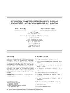
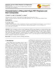
7 Following chemotherapy, whole body MIP image obtained July 2007, (B) there has been a dramatic reduction in the uptake of FDG within the mediastinum and supraclavicular region, but there is increased uptake within the bone marrow that is related to increased hematopoietic activity as a response to chemotherapy Brazilian Archives of Biology and Technology Common Causes of False Positive F18 FDG PET/CT Scans in Oncology 31. B. A C. Figure 3 - This 6 year old child underwent PET/CT for evaluation of right supraclavicular lymphadenopathy. The MIP image (A and B) shows intense uptake of FDG in the bilateral supraclavicular regions that would be suspicious for disease, but when correlated with the fused image (C), this area of increased uptake is in the region of fat and is consistent with brown fat.
8 The CT portion of the exam helped exclude our original diagnosis of lymphoma that was obtained from interpreting only the PET. images DISCUSSION result in a False Positive diagnosis for malignancy on PET/CT . Several cases have been reported FDG is not only a cancer specific imaging agent, including abscesses (brain, abdominal, renal, tubo- False Positive results may be observed with benign ovarian), osteomyelitis, sinusitis, myositis, diseases. False Positive results are commonly thryoridits, matitis, esophagitis and gastritis (Alavi observed in areas of active inflammation or et al., 2002). Other described etiologies include infection (Gupta et al.)
9 , 20000), with a reported pneumonia from multiple Causes (Bunyaviroch False Positive rate of 13% and False negative rate and Coleman, 2005). While there are numerous of 9% (Alavi et al., 2002). Inflammatory cells etiologic Causes for infection, tuberculosis and the (neutrophils and activated macrophages) at the fungal infections (Cryptococcosis, Histoplasmosis, sites of inflammation or infection will show Coccidioidomycosis, Blastomycosis, and increased FDG accumulation (Alavi et al., 2002). Aspergillosis) are most commonly described as These False - Positive areas of metabolic activity source of False Positive results with PET/CT .
10 Have the potential for significant morbidity and examinations (Bunyaviroch and Coleman, 2005). mortality if not accurately recognized. Tuberculosis is one of the oldest diseases to affect Brown adipose tissue has been reported to be humans. It is caused by bacteria belonging to the observed in 2-4% of patients and is especially mycobacterium tuberculom complex (Raviglione Common to be observed in women and children and O'Brien, 2005). It usually affects the lungs, during cold weather months. This tissue is but in one third of cases, other organs are responsible for cold induced and diet induced involved. Extrapulmonary sites may also be thermogenesis.