Transcription of Companion animal fluid therapy part 2: planning and …
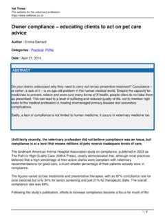
1 Vet Times The website for the veterinary profession Companion animal fluid therapy part 2: planning and monitoring Author : Rebecca Robinson Categories : Companion animal , Vets Date : September 19, 2016. Part one of this article ( ) introduced fluid therapy , fluid dynamics and the variety of fluid types available. Such knowledge is a prerequisite to safely and effectively develop fluid therapy plans, which are discussed in this part. Approaches to fluid therapy Table 1. Clinical findings that can indicate the degree of dehydration present. Animals may require fluid therapy for numerous reasons, including restoration of intravascular volume, correction of dehydration, treatment of electrolyte disturbances (beyond the scope of this article) or simply when the animal is not able to match its daily fluid requirements with adequate food and/or water intake.
2 When devising a fluid therapy plan, consider the following questions (DiBartola and Bateman, 2012): 1 / 16. Does the animal need fluid therapy ? What type of fluid needs to be given? What route of administration should be used? How much fluid should be given? How rapidly should the fluid be given? When should fluid therapy be discontinued? Assessment of fluid therapy requirement A thorough history and clinical assessment is a critical starting point to determine how much and what type of fluid is required. An accurate history can provide clues to the chronicity of the process, along with the type of body fluid lost.
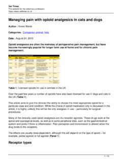
3 A detailed clinical examination especially of the cardiovascular, respiratory and neurological systems is vital. fluid loss can manifest as either hypovolaemia or dehydration. This depends on both the speed of fluid loss and the compartment the fluid has been lost from: Hypovolaemia reduced intravascular volume occurring with plasma water or whole blood loss. Dehydration a water deficit associated with all fluid compartments, thus causing an overall increase in electrolyte concentrations. Table 2. Clinical findings that can indicate the severity of hypovolaemia present. These two states cause different clinical signs.
4 It is important to be able to differentiate between dehydration (Table 1; Goggs et al, 2008) and hypovolaemia (Table 2; Boag and Hughes, 2007) as the treatment plan will vary. A patient with acute, severe hypovolaemia may present in shock. In these situations it is critical to differentiate hypovolaemic shock from cardiogenic shock (Goggs et al, 2008), which occurs due to cardiac pump failure. The fluid required for restoration of intravascular volume in hypovolaemic shock can be potentially fatal in cardiogenic shock. Discrimination between the two can be made by a patient's history, signalment and clinical findings (presence of pulmonary oedema, for example).
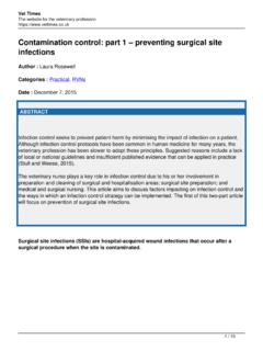
5 2 / 16. fluid therapy required Panel 1. Example of potassium addition. The details of additives should always be written on the fluid bag, as this will often be specific for the individual patient. It also helps prevent inadvertent fluid boluses from potassium-supplemented fluids being administered Body mass of dog: 25kg. Required infusion rate of fluids: 4ml/kg/hr. Serum potassium level: Amount of potassium to add to 250ml NaCl: 15mmol. fluid type required: Hartmann's solution. Amount of potassium contained in Hartmann's solution: 5mmol/L. Size of fluid bag: 1,000ml. Total amount of potassium to add: 55mmol (15mmol per 250ml = 60mmol per 1,000ml - 5mmol/L contained in Hartmann's solution).
6 Concentration of potassium solution: 13mmol per 5ml ? Volume of potassium to be added to fluid bag: Final concentration of potassium in fluids: 60mmol/L. ? Infusion rate of fluids: 100ml/hr. Total potassium infused per kg, per hour: (this is a safe infusion rate of the potassium containing fluids, as it is below the stipulated ). Different fluid types were considered in detail in part one of this article. Broadly speaking, most patients requiring fluid therapy can be managed with a small selection of crystalloid solutions and additives (DiBartola and Bateman, 2012). The most useful are balanced extracellular fluid replacers, which have a composition similar to plasma (see Table 4 from part one).
7 The most commonly used commercially available solution in the UK is Hartmann's solution, with Plasma-Lyte being more common in the US. As discussed in part one, even these solutions are not ideal for prolonged use. Compared to an animal 's electrolyte maintenance requirement, they are low in potassium and high in sodium and 3 / 16. chloride. This can result in hypokalaemia with a concurrent hyperchloraemic metabolic acidosis. Vets often rely on a patient's kidneys to resolve any iatrogenic disturbances, although this could become a problem in those with compromised renal function. Colloids are usually reserved for use in one of three situations.
8 Cases of hypoproteinaemia (most commonly hypoalbuminaemia) when colloid osmotic pressure (COP) is reduced. Use of colloidal solutions in this situation can boost COP, aiding with maintenance of intravascular fluid . When rapid intravascular expansion is required, particularly when large volumes of fluids passing into the interstitium (causing oedema) may be contraindicated. Colloid solutions do not redistribute as rapidly, so give more effective intravascular volume expansion for a smaller volume administered compared to crystalloid solutions. When a longer duration of effect is required (compared to crystalloid solutions, which rapidly redistribute).
9 Although, in most cases, use of balanced extracellular fluid replacers is suitable, the vet should aim to replace losses with a fluid similar in volume and electrolyte composition to that lost (DiBartola and Bateman, 2012). One rare example of a clinical indication for administration of a solution other than a balanced electrolyte crystalloid is persistent vomiting. Such patients lose stomach hydrochloric acid through vomiting, as well as potassium and water from lack of intake. Metabolic alkalosis may also occur due to the loss of acid. Potassium-supplemented per cent NaCl will help replace that lost. Administration of per cent NaCl also has a mild acidifying effect.
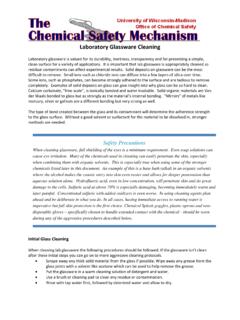
10 Additives Table 3. Amount of supplemented potassium required depending on serum potassium levels. 4 / 16. Absolute rate of potassium administration should not exceed As many commercially available fluids do not fit the exact specifications required, it is common to add other substances before administration. The most common additive is potassium chloride (KCl). Many hospitalised patients are hypokalaemic, either as a direct result of underlying disease processes, anorexia or fluid therapy . Addition of KCl helps counteract the severity of hypokalaemia, which may ensue with mid-to-long- term use of the commonly used extracellular fluid replacers.