Transcription of Compound odontoma – diagnosis and treatment: three case ...
1 Pediatric Dentistry 23:2, 2001 American Academy of Pediatric Dentistry 151 Oral PathologyCompound odontoma diagnosis and treatment: three case reportsBranca Helo sa de Oliveira DDS, MS Vera Campos DDS, MS Sonia Mar al DDS, MSDr. Oliveira and Dr. Campos are assistant professors, and Dr. Mar al is associate professor, Department of Community andPreventive Dentistry, Faculty of Dentistry, Rio de Janeiro State University, Brazil. Correspond with Dr. Oliveira are the most common type of odontogenic tumorsand generally they are asymptomatic. This paper describes threecases of Compound odontomas diagnosed in children due todislodgement or over-retention of primary anterior teeth and/orswelling of the cortical bone.
2 In all cases, the surgical excision ofthe lesions was performed and, in one case, orthodontic treatmentwas adopted in order to move an impacted permanent tooth to itsnormal position. The results achieved indicate that the early di-agnosis of odontomas allows the adoption of a less complex andexpensive treatment and ensures better prognosis. (Pediatr Dent23:151-157, 2001)Odontomas are considered to be developmental anoma-lies resulting from the growth of completely differen-tiated epithelial and mesenchymal cells that give riseto ameloblasts and odontoblasts. These tumors are basicallyformed of enamel and dentin but they can also have variablesamounts of cement and pulp During the developmentof the tumor, enamel and dentin can be deposited in such away that the resulting structures show an anatomic similarityto normal teeth, in which case the lesion is classified as a com-pound odontoma .
3 However, when the dental tissues form asimple irregular mass occurring in a disorderly pattern, it isdescribed as a complex Compound odontomas ap-pear more frequently than complex ,4 These odontogenic tumors can be found anywhere in thedental arches. The majority of odontomas which are locatedin the anterior region of the maxilla are Compound , while thegreat majority of odontomas located in the posterior areas, es-pecially in the mandible, are complex ,5,6 The etiology of the odontoma is However, it hasbeen suggested that trauma and infection at the place of theMajor CharacteristicsCompound OdontomaComplex OdontomaFrequencyThe relative frequency amongThe relative frequency amongodontogenic tumors varies betweenodontogenic tumors varies9% and 37%.
4 It s considered thebetween 5% and 30%.commonest odontogenic majority of cases appear beforeThe majority of cases occur beforethe age of 20, making it a lesion ofthe age of 30 with a peak in thechildhood/adolescence. second decade of and female subjects areMale and female subjects areequally anterior regionPosterior mandibular followedis the most frequent anterior maxilla are themost frequent presentationPainless, non-aggressive lesion, withPainless, slow-growing anda more limited potential growth thanexpanding lesion. Oftenthe complex odontoma . Oftenassociated with an uneruptedassociated with an uneruptedpermanent featuresRadiopaque mass of multiple, small,More or less amorphous mass ofcalcified structures with an anatomicalcalcified material with the radiodensitysimilarity to normal teeth usuallyof tooth structure, which bears nosurrounded by a narrow radiolucent resemblance to tooth,surrounded by a narrow radiolucent surgical surgical 1.
5 Major Characteristics of Compound and Complex OdontomasReceived May 18, 2000 Revision Accepted November 18, 2001152 American Academy of Pediatric DentistryPediatric Dentistry 23:2, 2001lesion can offer ideal conditions for its ,8 In gen-eral they are asymptomatic, have slow growth,1 and seldomexceed the size of a tooth, but when large can cause expansionof the cortical ,2 Odontomas may be diagnosed at any age but they are usu-ally detected during the first two decades of ,3 One studyanalyzed 396 cases and showed that diagnosis usually happensbetween ages 11 and 15 Another study comprising 149cases concluded that the lesions are detected most often dur-ing the second decade of Many times odontomas are foundassociated with unerupted ,6,9,10-14 The canines.
6 Followedby upper central incisors and third molars, are the most fre-quent teeth impacted by In a very few instancesodontomas are related to missing Generally these mal-formations are intraosseous, but occasionally they may eruptinto the oral ,14 Radiographic aspects of odontoma are characteristic. Thecomplex odontoma appears as an irregular mass of calcifiedmaterial surrounded by a thin radiolucent area with smoothperiphery, and the Compound type shows calcified structuresresembling teeth in the center of a well-defined radiolucentlesion. A periodontal and pericoronary space characteristic ofunerupted teeth is seen around each ,7 A developingodontoma can be discovered by routine radiography, but maycause difficulty in identification due to lack of histological examination of odontomas often shows thepresence of enamel matrix, dentin, pulp tissue, and cementumthat can, but need not, exhibit a normal ,7 Com-pound odontomas are formed by tooth-like structures whichresemble pulp tissue in the central portion surrounded by adentin shell and partially covered by enamel.
7 Complex odon-tomas are conglomerates without orientation of dentin, enamel,enamel matrix, cementum, and areas of pulp tissue. The cap-sule of connective tissue that surrounds an odontoma is similarto the follicle that covers a normal are treated by conservative surgical removal andthere is little probability of ,8 Ameloblastic fibro-odontomas and odontoameloblastomas show a greatresemblance to common odontomas, especially in the radio-graphic examination. Therefore, it has been suggested that allspecimens should be sent to an oral pathologist for ,8 Besides, proper patient care should includecareful clinical and radiographical ameloblastic fibro- odontoma is defined as a tumor withthe general features of an ameloblastic fibroma but that alsocontains enamel and ,16 It is usually encountered inchildren with an average age of 10 It has been suggestedthat ameloblastic fibro-odontomas should not be consideredas true neoplastic odontogenic lesions.
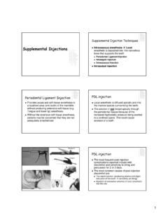
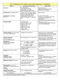
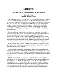
8 But rather as a stagepreceding the complex odontoma which would be the finalFig 1. Case 1. Clinical aspect showing dislodgement of the upper leftprimary central 2. Case 1. Periapical radiograph showing two lesions of 3. Case 1. three mineralized structures removed from the interior ofthe Dentistry 23:2, 2001 American Academy of Pediatric Dentistry 153stage in this line of development of hamartomatous authors consider that, despite the fact that some lesionsdiagnosed as ameloblastic fibro-odontomas can be developingodontomas, all cases of ameloblastic fibro-odontomas shouldnot be considered as hamartomatous in nature since there arerare cases of ameloblastic fibro-odontomas showing true neo-plastic The majority of the ameloblasticfibro-odontomas are found in the posterior region of the man-dible.
9 These lesions seem to be exclusively central orintraosseous tumors. They are also characterized by being pain-less and Radiographically, the tumor shows awell-defined unilocular or, rarely, multilocular radiolucentdefect that contains a variable amount of calcified material withthe radiodensity of dental hard tissues. The calcified materialwithin the lesion may appear as multiple, small radiopacitiesor as a solid conglomerate ,15 It can be differentiated fromthe odontoameloblastoma by the fact that it is well circum-scribed and usually separates easily from its bony Thetreatment of choice is conservative surgical enucleation andprognosis is ,15 However.
10 Development of anameloblastic fibrosarcoma after curettage of an ameloblasticfibro- odontoma has been ameloblastic fibrosarcoma is considered to be the ma-lignant counterpart of the ameloblastic fibroma and oftenrepresents a recurrence of a tumor previously diagnosed as anameloblastic fibroma or an ameloblastic Itis characterized by a malignant transformation of theectomesenchymal component of the tumor and not the odon-togenic The average age at time of diagnosis forthe ameloblastic fibrosarcoma is years, as opposed to for the ameloblastic fibroma and 9 years for theameloblastic fibro- odontoma .