Transcription of COTTONOSTEOTOMY:TechniqueandComplications
1 INTRODUCTIONThe Cot ton oste otomyis a medialcune iform openi ngwed ge oste otomywith the wedg e base d dors al ly topl ant ar flex th e media l co lu mn (1). It has been trad iti onall yus ed in co nju nctio n with oth er pro cedu res to co rrect a pe spl anovalgus foot wi th forefoot va rus defo rm it y. The authormost comm only uses the proc edure as an adjunctto anEva ns ca lcan eal os teoto my and ga str ocn emiu s rec essi on forcorr ect ion of a flexib le pedi atric pes pl ano valg us de for Cot ton oste otomy has additi onall ybeen use dfor hall uxlimi tus , me ta ta rsus primusel eva tus, as an add itiona lproc edurein adult flatfootcorr ec tion, and for efoo tsupinatus. Al ter na ti ves to thi s pr oc edur e inclu de any varie tyof medial co lumn fusion s suc h as a na viculo cun eifo rm joi ntar thr ode sis or fir st met at ar soc uneiform joint art hr odesi s,a plant arf lex ory first met atar sa l osteotomy, or soft ti ssu emedia l arc h rec ons tructio nt ag es to th e Cot ton ost eoto my co mparedto thelat ter li st of pro cedu res is it s si mpli cit y, reliableresul ts, easeof intr aope ra tive adjust ab ili ty, and pr eser vat ion of themedi al columnjoints (2, 3).
2 In addit ion, the Co tt onos teot omy len gthe ns the medi al col um n as opposed to theshort eni ng tha tocc urs typica lly wi th arth ro des is proced ures .There are few doc umentedcompl ic ati on s with th e Co ttonos teot omy, an dth eau thor s own experience co rr es pon ds toth is .Howe ver ,the author has exp erienced afew in sta nces ofcompl ic at ion s in th e use of this proc edu re. Thes e instancesas we ll as pre viouslydocu mentedcom plica tions wi ll bere view ed in th is up proc edure is perfor med with the pat ie nt in a supi nepos it ion. Typ ical ly, a posteriorlengthening(gast ro cne miusreces si on or tendo -Achilleslen gth en in g) and the primar ycorr ect ive pr ocedure(Evanscal caneal osteot omy, subtalarjoint fus ion , etc.) are perfo rmed first. The su rg eon will the npl ac ethe su btalar joi nt in aneut ra lposit io nan deval uate themedia l co lumn stabili ty and posit io n.
3 On ce th e sur ge on hasdeci ded to corr ect any res idual forefootvarus wit h th eCott on oste otomy , the inc is ion is vari ety ofinci sion plac emen ts ha ve been desc rib ed in clu din game dial ,dorso medi al, or dorsal linear incis ion ove rlying the medi alcun eifo rm. The au thor s pre fe ren ce is to make a lineardo rsal to dor som ed ial in ci si on 3-4 cm in len gth over lyi ngthe me di al cune if or m. Ana tom ic di ssec ti on is perf or med toavoi d the me di al dorsa l cu ta neo us nerve and th e sa phe nous(m edi al margin al ) ve in and ner ve . Perf oratorsof the me di alma rgi na l ve in ma y be en co unte re d and ligate d as ne cessaryand th en the me dia l ma rgi nal vei n is re trac ted late ral ly (4).A lin ear cap su la r-p er io ste al in ci sio n just med ial to th eext enso r hal luci s long us te ndon is mad e wit h care no t todisr upt th e dorsa l li ga me nts of the adjace nt joint s and thetibia lis ante ri or tendontha twill be jus tmedia land pla ntar tothe pl anned oste oto my (2).
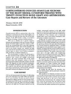
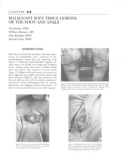
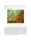
4 A va rie ty of method s can be uti li zed to ident if y the firstmeta tars ocun ei fo rm an dnavi cu loc un ei fo rm joints to ensureth at th e ost eo to my wi ll be extra-art ic ul ar. Th ese in cl udepa lpatio nof the se jo in ts an dthe sec on dmetata rsocuneiformjoi nt, use of a25 or 27 ga ug ene edleinser te dint oth ejoi nts,and int raop erat ive C- ar m flu or osco py. The le vel of theost eot om y is best place d, in th e aut ho r s opi nion , justpr ox imal to the levelofth ese con dme tat arsocu nei form thou gh ot he rs hav e sug gest ed th at th e seco ndme ta ta rso cune if orm joi nt le vel allow s easie r mobil iz at ion ofth e oste oto my, on e of the com pli cat ions the auth or hasexp eri ence d is th at of in te rp os it io n of the graft int o th ese con d metat ars ocu ne if or m joi nt req ui ri ng su rg icalre-i nt er vent ion . Th e tra ns verse ost eot om y is tha n crea tedus ing asagi ttal saw from dor sa lto planta rpara lleling the fir stme ta ta rso cune if orm joi nt (not para ll el to the we ig htbe ari ngsu rf ac e) while ta kingcare to pres erve th emedial cuneif or m spl an ta r corte x (Figu re 1).
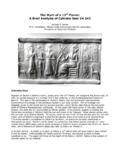
5 Com pl et evisualizat ion of the medial and lat eral edg es ofth e me di al cun eifo rm ar e necessa ry to ensu re both th eme dial and lat eral co rt ic es are cu t pri or to at temp tedop eni ng of the ost eot om y (Fi gu re 2). If one of theseco rtic es fa il to be cut, tha n the pla nta r hi nge may fra ctu rein to th e prox imal or di st al joi nt when the os teot om y isope ned (4). Onc e th e cut is pe rf orm ed, the osteot omy canth en be ope ned a var ie ty of way s in clu di ng osteo to mes, aSy nthe s mini -di stractor, a We inr aub dist ract or (I nnome d,Sa vanna h, GA) , or smoo th la mi na spr eade r (2) . The autho rpre fe rs us in g eit he r the mini -di stract or or Wei nrau bdist ractor , which all ows pi n place ment di stal and pro xim alto th e ost eot omy. Thi s all ow s adju st abili ty of distraction toin tr aop erat ively de te rmin e th e amo un t of desiredCOTTON OSTEOT OMY:Technique and ComplicationsAnnette li at rault,DPMCHAPTER32 CHAPTER32167pl an tar fl ex ion for deformity correc tion and it off er s anun obst ruc ted view for mea surementof the desir ed graftwid th/ di mensi on s on ce th at ha s been det ermin ed.
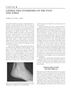
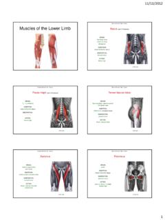
6 It alsoallowsan ea sier insertionof the gra ft by openi ng theost eoto my slightlygreat er tha n the pl annedgraf t uponinser tion. The dim ensio ns ofth egr aft deter mine the amo untof pla nta rflexi on achi e gener al id ea is to re-est ab li sh wei ghtbearingof th efi rst met ata rsa l he ad equal to the fi fth met at ars al head asdic tate dby Co tton sconcep tof the foot as atri pod (1). Thegra ft is ta mpe d into the osteotomywith care taken to noten ter the se cond metatarsoc unei for m joint or bur y thedor sal cor tex of the gra ft past the dorsal edg e of the me dialcun ei form (Figure3). The author typ icall y uses a fr eez e-dri ed il iac cre st wedgeallog ra ft (usua lly le ft over fr oman Eva ns proce dure) cut toth edes ire ddimens ions (typic ally4-8 mm at the dorsal wedg e bas e ta peringplanta rly in atrape zo ida l manne r) for correction wi th the cortexbaseddor sa ll y, alt houghautog raft coul d certainlybe used as th esurgeonfeels ap pr opri ate.
7 It ha s been the exp erienceof theauthoras well as others th at gra ft inc orp or at io n is ty pi cal lynot apro bl em wi th fr eeze-driedallograft(5) ,al th ou gh it hasbe en obse rv ed that time to incorp orationmay be pr ol on gedin adult pa tie nts (8-16 week s, avera ge 12) compa red topedia tri c patients (6-8 weeks)(6).Figure1. Dorsomedialskin and periostealincisionshave been performedexposingthe dorsal surfaceofthe transverselines on theskin were markedusing intraoperativefluoroscopyat the proximaland distal adjacentjoints to helporient the 2. The completedosteotomywith care takento cut both the medialand lateral corticeswhilemaintainingthe The osteotomywas openedwithosteotomesand the graft impactedinto 32 The gr aft may be remo deled after pl acem ent as neede ically ,th egraf tis dri ven from adors alto pl anta rdirect ionfor pl ant arfl exion , ho wever, it may be dr iven dorsome dial lyas an atte mpt to providesom e tr ans ve rse int erm etat arsa lco rre cti on for a bunionas wel l as plant arfl exio n (4).
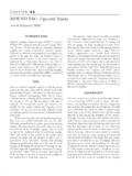
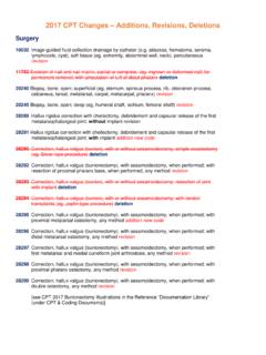
8 Fixati on is typ ical ly no t necessa ry if the gr aft press fi ts we llin to the osteotomy site . Opt ions fo r fixa ti on, if desired bythe surg eon, cou ld includea Ki rschne r wire, stap le, scre w,or two- or thr ee-h ole pl ate. On ly when th e gr af t does no tappear to ti gh tl y oppose the ad ja cen t bon e has the auth orused fi xa tion (F igure4). The aut hor rec om mendsint raop er ativ e fluoroscopyto revi ew graft plac eme nt priorto soft ti ss ue clo su re, wh ich is perfo rmed in layer s. A no n-wei ghtbe aring be low- knee cas t is app lie d postoperativel yunt il time of gr aft incorporation, whi ch as note dearlie r, maybe longer in ad ults .COMPLICATIONSCo mp lica tio ns can result from su rgeo nerror per fo rmi ng thepr oceduresuch as pr oper pa ti ent selec tio n, join t viol ation ,und ercorrection with lateral foot pa in , overco rrecti on withsesa moid pain or plantarfasci iti s, and dis se ct ion-re late dsuchassaphen ou svein ornerve disrupti on or ten do ndi srup ti yed unionis com mon wi th the adult popul at io nco mpare d to the pediat ric popul ati on, but nonunion hasrare ly be enreport ed (4 ,6).
9 Other compli cat ions can includebo ne gr aft displ acementeithe r dors ally or in to the seco ndmet ata rs ocune ifor mjoint and bo ne graft col lapse with los sofcorr ect ion , whic h are also rar ati vel y fe w comp lic ati ons have been documentedwit h th e Cottonproc edur e or ex peri enc ed by the author;howe ver th eau tho rhas experienced afew co mplicationsth atserv e to emp has ize the import anc e of ea ch su rg ical step inthe above descri bed proc ed ure .Thefir st ca se was apedi atr icpe s pla no valg us co rr ection in wh ic h the Cotto n osteo tomywas cr eat ed more perpendicular to the foot th an theor ient at ion of the fi rst meta ta rsoc unei for m join t, ther eforethe ost eo to my cam e close to ent eri ng th e in ferio r asp ect ofthe joi nt. Th e pa ti ent ha d som e tra nsient ten dern ess to thisar ea po stope rat iv ely, but this eventuallyre sol ved wi thorthot ic s.
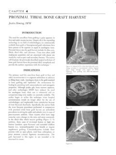
10 The author now doubl e che cks the pro po sedorie nt at ion of the osteo tomy an d lo cation of the adjac entjoint s wit h C-ar m fluo rosco py prio r to the per for manceofthe ost eotom y. The secondca se wa s also a pe dia tri c pespl an oval gus cor re ction in whic h the graf t on the ini ti alpos tope rative films appeared to be wel we ekspos tope rativel y, the patien t report ed a fall in th e bathr oomand ra di ographsde monst rated inter pos it ion of the graft in tothe se cond met at ar socuneiform joi nt (F igur e 5). Thi spat ie nt wa s take n back to surger y that day, the graf t wasre pos itione d wit h little di ffic ul ty , an d the graft ul ti mat elyin cor po rat ed un ev entfu lly. Last ly, a thi rd pat ien t fo rped ia tri c pes pl an ov al gu s co rrect ion was noted to havein te rp osi tion of the gra ft in to th e secon d met atars o-cu neifo rm join t on in tr a- oper at ive flu orosc opy (F igure 6).