Transcription of DCM01(obsolete) DCM02 DCM03 DCM05 DCM06 DCM07 …
1 dcm01 ( obsolete ) DCM02 DCM03 DCM05 DCM06 DCM07 DCM08 DCM10 To measure arterial oxygen saturation, when pulse oximeters with transmission probes cannot be used, pulse oximeters with reflectance probes are used to SpO2 based on the intensity of reflected light. The idea of using light reflection instead of light transmission in clinical oximetry shows that SpO2 can be monitored by measuring the amount of light reflected from the tissue. The idea of using skin reflectance spectrophotometry marked a significant advancement in the noninvasive monitoring of SpO2 from virtually any point on the skin surface. Eventhough it was a major advancement, difficulties in absolute calibration and limited accuracy were the major problems with early reflectance pulse oximeters. dcm01 , DCM02 , DCM03 , DCM05 , DCM06 , DCM07 , DCM08, DCM10 developed by APMK orea is a sensor integrated LEDs and PD together, assuring the accuracy and minimizing any variance to get reliable parameters.
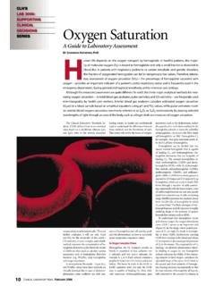
2 The intensity of the back scattered light from the skin depends not only on the optical absorption spectrum of the blood but also on the structure and pigmentation of the skin. SpO2 is measured be analyzing the pulsatile componens of the detected red and infrared plethysmograms which make use of reflected light intensities. The light from the LEDs enters the tissue, is scattered by both the moving red blood cells and the nonmoving tissue, and a part of this back scattered light is detected by the photodiode. The output of photodiode is processed by the pulse oximeter , and measures the SpO2 of the pulsatile blood. In reflectance pulse oximetry, the LEDs and the photodiode are placed on the same side of the skin surface as shown below. Normally the reflectance probe is placed on the forehead or temple, but is not restricted to only those two places.
3 Reflectance probes can be used to measure arterial oxygen saturation at virtually any place the human body where the probe can be placed. DCMXX Reflected light One of the major design considerations required in designing a reflectance pulse oximeter sensor is determining the optimum separation distance between the LEDs and the photodiode. This distance should be such that plethysmograms with both maximum and minimum pulsatile components can be detected. These pulsatile components depend not only on the amount of aterial blood in the illuminated tissue, but also on the systolic blood pulse in the peripheral vascular bed. There are two techniques that can enhance the quality of the plethysmogram. ( ) One way is to use a large LED driving current, which determines the effective penetration depth of the incident light, which increases light intensity.
4 So far a given LED/photodiode separation, using higher levels of incident light, you can illuminate a larger pulsatile vascular bed. As a result the reflected the plethysmograms will contain a larger AC components. But in practice, the LED driving current is limited by manufacturer to a specified maximum power dissipation. The other way is to place the photodiode close to the LEDs. If you place the photodiode too close to the LEDs, the photodiode will be saturated as a result of the large DC component obtained by multiple scattering of the incident photons by the blood-free stratum corneum and epidermal layers in the skin. For a constant LED intensity the light detected by the photodiode (AC & DC) decreases roughly exponentially as the radial distance between the LEDs and the photodiode is increased. In other hands, the effect of LED/PD separation on the relative pulse amplitude of both red and IR plethysmograms is increased with the increase in separation.
5 Thus the selection of a particular separation distance involves a trade-off. You can achieve larger plethysmograms by placing the PD farther apart from the LEDs but you need higher LED driving currents to overcome absorption due to increased optical path length. DCMXX developed by APMK orea is a sensor integrated built in LEDs and PD together with optimized properly against such trade-off to get proper parameters. skin blood electrodes Furthermore dcm01 dcm01 , DCM02 , DCM03 , DCM05 , DCM06 , DCM07 , DCM08, DCM10 has SMD type package so that user could interconnect easily wire or flip bonding into your set. DCM04 has green and IR combo Leds, not for SpO2 but different wavelength 570nm, DCM05 is smallest among DCM family which is suitable for mobile devices. This reflectance sensor can be placed on the body where you can expect light reflection due to tissue.
6