Transcription of Dentigerous Cysts But what An Avoidable …
1 From The CUSP, January 2007 Page 1 Fraser A. Hale, DVM, FAVD, Dipl AVDC Dentigerous Cysts An Avoidable catastrophe Some dental problems just happen despite all best efforts. Labs will break teeth, border collies will wear their teeth down, toy poodles will get periodontal disease, Shih Tzus will suffer mandibular fractures as a result of being out in a strong wind. There is one problem, however, that is 100% Avoidable ; and avoid it we must. This would be the Dentigerous cyst . By definition, a Dentigerous cyst (in a dog or cat) is a fluid-filled cyst surrounding the crown of an unerupted tooth.
2 This is in contrast to the equine version, which I will not be discussing. It is time for some background. When a tooth is in the early developmental stages, there is a multilayered sac of tissue surrounding the primordial dental pulp. This sac, known as the enamel organ, covers the pulp like a toque* without the pompom. The outer layer is the outer enamel epithelium. Inside this is the stellate reticulum. The inner surface, in contact with the pulp, is the inner enamel epithelium, made up of the ameleoblasts.
3 These are the cells responsible for the production and mineralization of the enamel that covers the crown of the tooth. Once the coronal enamel has been formed, the enamel organ has out-lived its purpose and it shuts down. The stellate reticulum atrophies and the outer enamel epithelium collapses onto the inner enamel epithelium. At this point, the structure becomes the reduced enamel epithelium. When the tooth erupts and the crown breaks through the gingiva, most of the outer enamel epithelium is torn away.
4 A small ring of it persists near the junction of the crown and root to form part of the gingival attachment (the junctional epithelium). It is no longer a closed sac, but a collar of tissue surrounding the neck of the tooth. But what happens if the tooth fails to erupt? sometimes nothing happens. sometimes the tooth and its surrounding tissues just lie dormant in the bone. sometimes the tooth is absorbed. sometimes something far worse happens. As cells from the inner enamel epithelium desquamate into the lumen of the sac, an osmotic gradient develops and fluid is pulled in, causing the sac to expand.
5 As it expands, it causes pressure atrophy of the surrounding bone. Left alone, these Cysts will just keep expanding indefinitely, destroying everything in their way. Within a very short time, they can destroy the support for adjacent teeth and weaken the jaw to the brink of pathological fracture. They can also cause external resorption of the roots they come into contact with. By the time these Cysts grow large enough to be detected by the owner or by the naked-eye on clinical examination, much irreversible damage is done and the necessary surgery can be quite involved.
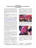
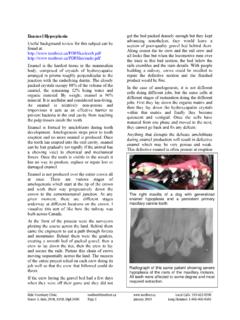
6 Specifically, it involves removal of the offending unerupted tooth, any adjacent teeth that have lost their periodontal support or have other radicular pathology and removal of the cyst lining from what is often a complex and convoluted cavern. Here is a clinical photo of a typical cyst in a young boxer. Note the definite blue hue to the overlying soft tissue. (If you are reading the B&W hardcopy of The CUSP, go to to view the full-colour pdf version.) This cyst has broken through the bone and the bluish area would be flocculent.
7 The good news is that Dentigerous Cysts are completely preventable and once you have read this article, you will never let one happen again. As is so often the case, the toque, n. 1 /tu:k/ Cdn a a close-fitting knitted hat, often with a tassel or pompom on the crown. From The CUSP, January 2007 Page 2 Fraser A. Hale, DVM, FAVD, Dipl AVDC key lies in a) knowing normal anatomy, b) looking carefully for abnormalities and c, d, e and f) taking intra-oral dental radiographs when there is even the slightest doubt.
8 If the radiographs reveal an unerupted tooth, get it out before a cyst has a chance to form. Problem averted and you are a hero because you just saved your client hundreds of dollars. Here s the deal. When you have a patient in at about six-months of age for spay/neuter always do a through oral inventory. There are many problems you might find and manage by routinely doing this, but let s stick to Dentigerous Cysts for now. A cat is supposed to have 30 adult teeth. A dog is supposed to have 42.
9 If you find less than this number, something is amiss. Maybe the adult teeth in question are actually missing and this requires only that you record the findings in the permanent record so you never have to wonder about it (them) again. On the other hand, maybe that missing tooth is not missing at all but is hiding (impacted or unerupted). If the tooth is unerupted there are a few options to consider depending on which tooth it is, where it is located, what direction it is pointing in and how far along the patient is in its oral development.
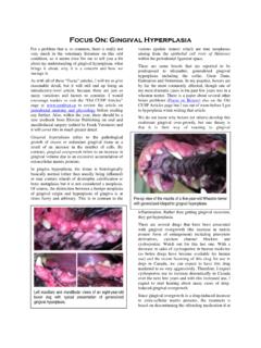
10 In a previous issue I discussed soft tissue impactions and how to deal with them. In those cases, the teeth have a good chance of erupting normally with a little surgical help (see ). In this piece I would like to consider a few examples of teeth that are not going to be able to erupt no matter what we do. You will then be able to extrapolate to the various clinical presentations you come across. In the image at the bottom of the last column, we see three abnormalities of note as well as some other useful information about this patient.