Transcription of Department of Dermatology, Rita Skin Foundation, Kolkata ...
1 Our Dermatology Online Letter to the Editor Striae distensae over scalp: A trichoscopic revelation Subrata Malakar, Priya Diwaker Department of Dermatology, Rita skin Foundation, Kolkata , India Corresponding author: Dr. Priya Diwaker, E-mail: Sir, also demonstrated linear, atrophic bands on her scalp;. striae distensae along with telangiectasia (Fig. 1). On Striae distensae is a common disfiguring cutaneous taking a detailed history, the patient confessed to condition which is characterized by linear, smooth and applying topical steroid for a long duration. atrophic bands over the skin . It is a reflection of breaks in the connective tissue and occurs at the areas of Keeping this trichoscopic finding in mind we dermal damage. The formation of striae distensae has subsequently started looking for similar linear atrophic been attributed to underlying mast cell degranulation bands on scalp of patients who were treated with topical with subsequent damage of elastin and collagen [1].
2 Steroids. On one hand, striae can be the end result of various A 17-year old male having patchy hair loss for 6 months physiological conditions like pregnancy, adrenocortical who was applying a lotion, bought over the counter, excess and change in the body habitus, while on the reported to us. He complained of itching at the site other hand they are indicators of excessive topical of application of the lotion. Trichoscopic evaluation steroid use [2,3]. confirmed him to be a case of alopecia areata (Fig. 2). He also had features of steroid misuse like telangiectasia Topical corticosteroid is considered as the first line and linear white atrophic bands suggestive of striae therapy in treating conditions like alopecia areata distensae. The lotion was later on identified to be and psoriasis. While its appropriate and accurate use fluocinolone acetonide %. can provide satisfactory results, its over-usage can lead to inadvertent effects such as development of A 28-year old female presented with hair loss for two erythema, telangiectasia, acneiform eruptions, striae years.
3 She had observed widening of her central parting and eventually atrophy and scarring. The misuse of and thinning of her pony tail. She visited a general steroid on the face can be picked up easily by the practitioner who prescribed beclomethasone lotion, signs mentioned afore. However on the scalp, due to which she had been using for the past one year with the presence of hair, these signs can easily be missed. no significant improvement. After taking detailed The role of trichoscopy holds importance especially in history to rule out chronic telogen effluvium, we treating scalp lesions as these signs can be identified proceeded for trichoscopy and observed hair diameter much before they are appreciated clinically. variability and multiple vellus hair at the frontal region which confirmed the diagnosis of female pattern hair We herein report 3 cases of striae distensae over the loss.
4 Trichoscopy also demonstrated the presence of scalp detected incidentally while examining the scalp telangiectasia with few, linear, white streaks which of patients on topical steroids. were arranged in parallel to form a band indicating striae distensae. A 35-year old female presented with patchy hair loss and was applying topical beclomethasone prescribed We would like to highlight that a tool as simple by her previous Dermatologist. On trichoscopy, there as a dermoscope can help us identify the steroid was presence of exclamation mark hair and yellow dots overuse. Periodic monitoring of the patient on topical confirming the diagnosis of alopecia areata. Trichoscopy corticosteroids can reveal subtle changes of dermal How to cite this article: Malakar S, Diwaker P. Striae distensae over scalp: A trichoscopic revelation. Our Dermatol Online. 2018;9(2):227-228. Submission: ; Acceptance: DOI: Our Dermatol Online 227.
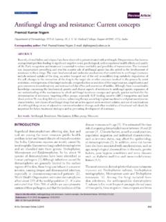
5 Damage. There might be a need to revise the treatment options in such cases and switch them to non-steroidal medications. An observant physician with minimal effort and time can easily recognize complications of steroid misuse like scarring and telangiectasia in patients and correct the damage done. Thus, trichoscopy is not only a diagnostic tool but also a monitoring tool wherein it can reveal the tell-tale signs of inadvertent topical therapy. Figure 1: Linear atrophic bands (straie distensae) and telangiectasia REFERENCES. over scalp on a patient with alopecia areata. 1. Sheu HM, Yu HS, Chang CH. Mast cell degranulation and elastolysis in the early stage of striae distensae. J Cutan Pathol. 1991;18:410-6. 2. Awley TJ, Yancey KB. skin changes and diseases in : Freedberg IM, Eisen AZ, Wolff K, Austen KF, Goldsmith LA, Katz SI, editors. Fitzpatrick's Dermatology in generalmedicine.
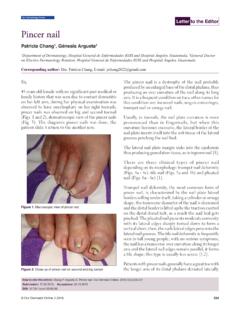
6 6th edn. New York: McGraw-Hill; 2003. p. 1362. 3. (Burrows NP, Lovell CR. Disorders of connective tissue. In: BurnsT, Breathnach S, Cox N, Griffith C, editors. Rook's Textbook of dermatology, 7th edn. Oxford: Blackwell Science; 2004. p. 46-7. Copyright by Subrata Malakar, et al. This is an open-access article distributed under the terms of the Creative Commons Attribution License, which permits unrestricted use, distribution, and reproduction in any medium, provided the original author and source are credited. Source of Support: Nil, Con ict of Interest: None declared. Figure 2: Telangiectasia over the scalp seen after steroid misuse. Our Dermatol Online 228.)