Transcription of Diagnosis and Management of G6PD Deficiency - American ...
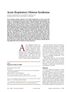
1 October 1, 2005 Volume 72, Number 7 American Family Physician 1277 Glucose-6-phosphate dehydroge-nase ( g6pd ) Deficiency increases the vulnerability of erythrocytes to oxidative stress. Clinical pre-sentations include acute hemolytic anemia, chronic hemolytic anemia, neonatal hyper-bilirubinemia, and an absence of clinical symptoms. The disease is rarely Deficiency occurs with increased fre-quency throughout Africa, Asia, the Mediter-ranean, and the Middle East. In the United States, black males are most commonly affected, with a prevalence of approxi-mately 10 percent. Prevalence of the Deficiency is correlated with the geographic distribu-tion of malaria, which has led to the theory that carriers of g6pd Deficiency may incur par-tial protection against malarial Cases of sporadic gene mutation occur in all catalyzes nicotinamide adenine dinu-cleotide phosphate (NADP) to its reduced form, NADPH, in the pentose phosphate pathway (Figure 14).
2 NADPH protects cells from oxidative damage. Because erythrocytes do not generate NADPH in any other way,3 they are more susceptible than other cells to destruction from oxidative stress. The level of g6pd activity in affected erythrocytes gener-ally is lower than in other Normal red blood cells that are not under oxidative stress generally exhibit g6pd activity at approxi-mately 2 percent of total Even with enzyme activity that is substantially reduced, there may be few or no clinical symptoms. A total Deficiency of g6pd is incompatible with The g6pd -deficient variants are grouped into different classes corresponding with disease severity (Table 11,7).GeneticsThe gene mutations affecting encoding of g6pd are found on the distal long arm of the X chromosome. More than 400 mutations Glucose-6-phosphate dehydrogenase Deficiency , the most common enzyme Deficiency world-wide, causes a spectrum of disease including neonatal hyperbilirubinemia, acute hemolysis, and chronic hemolysis.
3 Persons with this condition also may be asymptomatic. This X-linked inherited disorder most commonly affects persons of African, Asian, Mediterranean, or Middle-Eastern descent. Approximately 400 million people are affected worldwide. Homozygotes and heterozygotes can be symptomatic, although the disease typically is more severe in persons who are homozygous for the Deficiency . The conversion of nicotinamide adenine dinucleotide phosphate to its reduced form in erythrocytes is the basis of diagnostic testing for the Deficiency . This usually is done by fluorescent spot test. Different gene mutations cause different levels of enzyme Deficiency , with classes assigned to various degrees of Deficiency and disease manifesta-tion. Because acute hemolysis is caused by exposure to an oxidative stressor in the form of an infection, oxidative drug, or fava beans, treatment is geared toward avoidance of these and other stressors.
4 Acute hemolysis is self-limited, but in rare instances it can be severe enough to warrant a blood transfusion. Neonatal hyperbilirubinemia may require treatment with phototherapy or exchange transfusion to prevent kernicterus. The variant that causes chronic hemolysis is uncommon because it is related to sporadic gene mutation rather than the more common inher-ited gene mutation. (Am Fam Physician 2005;72:1277-82. Copyright 2005 American Academy of Family Physicians.) Diagnosis and Management of g6pd DeficiencyjENNIfEr E. frANk, MAj, MC, USA, Martin Army Community Hospital, Fort Benning, GeorgiaGlucose-6-phosphate dehydrogenase Deficiency occurs with increased fre-quency throughout Africa, Asia, the Mediterranean, and the Middle American Family Physician Volume 72, Number 7 October 1, 2005G6PD Deficiencyhave been identified, most being missense Most of the variants occur spo-radically, although the g6pd Mediterra-nean and the g6pd A variants occur with increased frequency in certain populations (Table 23,6,7).
5 DiagnosisThe Diagnosis of g6pd Deficiency is made by a quantitative spectrophotometric analysis or, more commonly, by a rapid fluorescent spot test detecting the generation of NADPH from The test is positive if the blood spot fails to fluoresce under ultraviolet In field research, where quick screening of a large number of patients is needed, other tests have been used; however, they require definitive testing to confirm an abnormal ,10 Tests based on polymerase chain reaction detect specific mutations and are used for population screening, family stud-ies, or prenatal patients with acute hemolysis, testing for g6pd Deficiency may be falsely negative because older erythrocytes with a higher enzyme Deficiency have been hemolyzed. Young erythrocytes and reticulocytes have normal or near-normal enzyme activity.
6 Female heterozygotes may be hard to diag-nose because of X-chromosome mosaicism leading to a partial Deficiency that will not be detected reliably with screening ,11,12G6PD Deficiency is one of a group of con-genital hemolytic anemias, and its Diagnosis should be considered in children with a family history of jaundice, anemia, spleno-megaly, or cholelithiasis, especially in those of Mediterranean or African should be considered in children and adults (especially males of African, Mediterranean, or Asian descent) with an acute hemolytic reaction caused by infec-tion, exposure to a known oxidative drug, or ingestion of fava rare, g6pd Deficiency should be considered as a cause of any chronic nonspherocytic hemolytic anemia across all population screening for g6pd Deficiency is not performed routinely in the United States, although it is done in countries with high disease prevalence.
7 The World Health Organization recommends screening all newborns in populations with a prevalence of 3 to 5 percent or more in : KEy REcOMMEnDATiOnS fOR PRAcTicEClinical recommendationEvidence ratingReferencesPatients with g6pd Deficiency should avoid exposure to oxidative drugs (Table 3) and ingestion of fava should be screened for g6pd Deficiency when family history, ethnic or geographic origin, or the timing of the appearance of neonatal jaundice suggests the possibility of g6pd can be diagnosed with a quantitative spectrophotometric analysis or, more commonly, by a rapid fluorescent spot 7 g6pd = glucose-6-phosphate = consistent, good-quality patient-oriented evidence; B = inconsistent or limited-quality patient-oriented evi-dence; C = consensus, disease-oriented evidence, usual practice, expert opinion, or case series. For information about the SORT evidence rating system, see page 1154 or Phosphate PathwayG6 PDHexokinaseATPNADP+ADPNADPHG lutathione reductaseGSSGGSHG lucoseGlucose 6 phosphate6 phospho- gluconatefigure 1.
8 g6pd catalyzes NADP+ to its reduced form, NADPH, in the pentose phosphate pathway. ( g6pd = glucose-6-phosphate dehydro-genase; ATP = adenosine triphosphate; ADP = adenosine diphosphate; NADP+ = nicotinamide adenine dinucleotide phosphate [oxidized form]; NADPH = reduced NADP; GSSG = oxidized glutathione; GSH = reduced glutathione.)Adapted with permission from Glucose 6 phosphate dehydrogenase Deficiency . Accessed online July 20, 2005, at: 1, 2005 Volume 72, Number 7 American Family Physician 1279G6PD Deficiencyneonatal HyperbilirubinemiaThe prevalence of neonatal hyperbilirubine-mia is twice that of the general population14 in males who carry the defective gene and in homozygous females. It rarely occurs in heterozygous ,16 The mechanism by which g6pd Deficiency causes neonatal hyperbilirubinemia is not completely understood.
9 Although hemoly-sis may be observed in neonates who have g6pd Deficiency and are jaundiced,17 other mechanisms appear to play a more impor-tant role in the development of ,18,19 Hyperbilirubinemia is likely secondary to impairment of bilirubin con-jugation and clearance by the liver leading to indirect ,20 Infants with g6pd Deficiency and a mutation of uridine diphosphoglucuronate glucuronosyltrans-ferase-1 gene promoter (UDPGT-1) are par-ticularly susceptible to hyperbilirubinemia secondary to decreased liver clearance of UDPGT-1 is the enzyme affected in Gilbert Deficiency should be considered in neonates who develop hyperbilirubine-mia within the first 24 hours of life, a his-tory of jaundice in a sibling, bilirubin levels greater than the 95th percentile, and in Asian ,23G6PD Deficiency can lead to an increased risk and earlier onset of hyperbilirubine-mia,24,25 which may require phototherapy or exchange ,25 In certain popu-lations, hyperbilirubinemia secondary to g6pd Deficiency results in an increased rate of kernicterus and death,26,27 whereas in other populations this has not been This may ref lect genetic muta-tions specific to different ethnic ,19 Acute HemolysisAcute hemolysis is caused by infection.
10 Inges-tion of fava beans, or exposure to an oxidative Medications that should be avoided in patients with g6pd Deficiency are listed in Table 3,6 and drugs that can be used safely in these patients are listed in Table Hemolysis occurs after exposure to the stressor but does not continue despite continued infection or ingestion. This is thought to be a result of older erythrocytes having the greatest enzyme Deficiency and undergoing hemolysis first. Once the population of deficient erythrocytes has been hemolyzed, younger erythrocytes TAble 1classes of g6pd Enzyme VariantsClass Level of deficiencyEnzyme activityPrevalenceISevereChronic nonspherocytic hemolytic anemia in the presence of normal erythrocyte functionUncommon; occurs across populationsIISevereLess than 10 percent of normalVaries; more common in Asian and Mediterranean populationsIIIM oderate10 to 60 percent of normal10 percent of black males in the United StatesIVMild to none60 to 150 percent of normalRareVNoneGreater than 150 percent of normalRareG6PD = glucose-6-phosphate from references 1 and 2 comparison of the Two Most c ommon Variants of g6pd Deficiency g6pd MediterraneanG6PD A World Health Organization classClass IIClass IIIP opulations affectedItalian, Grecian, Spanish, Arabic, Jewish (Kurdish)