Transcription of DIGESTIVE AND RESPIRATORY SYSTEMS - Biology
1 29 DIGESTIVE AND RESPIRATORY SYSTEMSThe DIGESTIVE system is responsible for obtaining and processing food for the all of the cells in anorganism. Structurally it is closely associated with the RESPIRATORY system so the two will be studiedtogether. Each system is composed of a series of tubular structures through which materials pass. In thecase of the DIGESTIVE system the raw materials are food particles that are broken into simple molecules asthey pass through the system . Material that is not broken down is removed from the system . In the caseof the RESPIRATORY system the major materials moving through it are oxygen and the waste product DIGESTIVE and RESPIRATORY system share some common spaces. The DIGESTIVE system is composed ofthe mouth, pharynx, esophagus, stomach, small intestine, large intestine, rectum and anus.
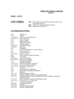
2 There are alsoa number of glands that lie outside the system that contribute to its function. These include the salivaryglands, liver and pancreas. The RESPIRATORY system is composed of the mouth or external nares, thepharynx, glottis, trachea, bronchi, bronchioles and alveoli. The latter three structures are in the lung. Asyou study these two SYSTEMS consider how the material moving through them is being glandsThree paired salivary glands lie in the head and neck region (Fig. ). Locate the following:Parotid gland. These triangular glands lie ventral tothe ear and extend along the lateral surface of the gland. These large oval glands cover theventral portion of the gland. These light colored glands areclosely associated with the mandibular ducts of the salivary glands enter the mouth.
3 Theglands produce mucus to lubricate food. In humans,they produce amylase for the breakdown of nodes . These are darker and less globularthan the salivary glands. They help initiate the immuneresponse and are the site of lymphocyte lacrimal gland. This gland lies cranial to the parotid. It is one of several glands thatproduce tears in the rat. The others lacrimal glands lie deeper in the and PharynxThe DIGESTIVE and RESPIRATORY SYSTEMS are closely associated in the mouth. Cut through the angle of thejaw on each side of the mouth. Push down on the jaw to expose the oral cavity. The tongue attaches atthe rear of the oral cavity. Lift the tongue and note that it is attached anteriorly by a sheet of tissueknown as the frenulum.
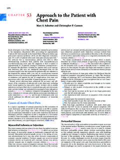
4 Also note the location of the two types of teeth, incisors for cutting and molarsfor grinding. The relationship between the spaces associated with the oral cavity can be confusing. TryFigure Glands of the neck the path food and air would take through this region(Fig. - ). Hard palate. The bony roof of the oral cavity. Soft palate. A continuation of the tissue lining the roof of the oral cavity. Pharynx. A space at the back of the oral cavity divided into: Nasopharynx. Dorsal to the soft palate the nasopharynxreceives air from the external nares and is not directly inthe oral cavity, Oropharynx. The space ventral to the soft palate. Laryngopharynx. The space posterior to the soft palateand anterior to the esophagus.
5 Both food and air passthrough the oral cavity, however their pathways divergein the laryngopharynx. Dorsally food moves into theesophagus and the DIGESTIVE system . Ventrally air movesinto the larynx and the RESPIRATORY (Fig. ) A slit-like opening into the larynx (1, ), leading via the trachea (2, Fig. ) into the lungs. Unlikethe esophagus, the trachea has rings of cartilage to keep it (Fig ) A flap of tissue that blocks the larynx when food or fluid is in the & Parathyroid glands (3, Fig ). Posterior to the larynx, paired thyroid glands controlmetabolism. A parathyroid gland is embedded at the anterior end of each. It regulates blood Cavities, pleura and PeritoneumThe body cavity of mammals is divided into an anteriorand posterior region by a muscular diaphragm.
6 Theanterior region is further divided into two lateral pleuralcavities that house the lungs and a central pericardialcavity that houses the heart. Posterior is the abdominal (peritoneal) cavity whichcontains the DIGESTIVE , reproductive and excretoryorgans. The walls of the body cavities and the surfacesof the organs are covered with an epithelium, derivedfrom mesoderm. Parietal epithelium lines the body walland covers the surface of the diaphragm. VisceralFigure Oral cavity. Inset: view into the slit-like glottis and triangular Sagittal section through the head of a ratshowing the path taken by air and Upper RESPIRATORY Sagittal section at the level of the covers the surface of the internal organs.
7 These two types of epithelium are further divided intopleura, pericardium or peritoneum depending on the body cavity in which they are found. Ventral to theheart the walls of the two plural cavities meet to form the mediastinal septum. The heart lies in a spacewithin the septum. The pericardial sac surrounding the heart is formed by the parietal pericardium andthe surface of the heart is covered with visceral pericardium (Fig. ).The abdominal cavity is lined with parietal peritoneum and the surface of the abdominal structures arecovered with visceral peritoneum. The organs in this region are attached to the body wall by extensionsformed by the passage of the peritoneum to and from the body wall. These extensions are known asmesenteries or ligaments.
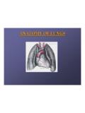
8 As you begin your dissection note the extent of the mesenteries and ligaments. They help keep the internalorgans in place. Also note that the majority of the mesenteries arise from the dorsal body wall. Opening the Body CavitiesTo open the body cavities you will need to make the five incisions shown in figure The skin shouldalready have been loosened on both sides along the midventral length of the body and into the groin area. Use care not to damage major blood centimeters to the right of the midline cut throughthe clavicle and posteriorly to the diaphragm. Repeat onthe left side using care not to damage the mediastinalseptum that divides the thoracic cavity into two laterally through the ribs and along the edge of thediaphragm toward the vertebral column.
9 Avoid cuttingthe diaphragm. a cut slightly to the right of the midline and followit posteriorly until just above the genital the skin has not been removed continue cut threelaterally through only the skin to the caudal margin of theischium. Use care so the underlying reproductivestructures are not the abdominal cavity cut laterally along the edge of thediaphragm toward the vertebral column. Spread the flaps of body wall to reveal the internal organs. Gently break the ribs near the spinal column to help hold thepleural cavity open. Carefully lift the midventral flap of skin and bone. Note themediastinal septum attached to this flap. It divides the thoraciccavity into two pleural Relationships between structures in the thoracic cavity.
10 Note the position of the multi- lobed lungs on either side of the heart and the position of the diaphragm andribs. The heart lies within the pericardial cavity formed from parietal pericardium (Fig. ). The surfaceof the heart is covered with visceral pericardium . Make sure you can distinguish between these two typesof pericardium . Also make sure that you can distinguish between the parietal and visceral pleura . SeeFigure for clarification of the relationship between the various types of Sequence of cuts for openingthe body - (Fig. ) this gland is part ofthe endocrine system . In young rats thethymus gland lies within the mediastinalseptum and over the anterior part of theheart. This gland is part of the immunesystem and functions in the maturation ofT-lymphocytes.