Transcription of Dilated Renal Pelvis - Kaiser Permanente
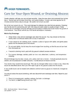
1 Renal pe vis Kidney bladder Dilated Renal Pelvis ~,,,~ Kaiser Permanente PRENATAL ULTRASOUND FINDINGS What is a Dilated Renal Pelvis ? Dilated Renal Pelvis A baby s kidneys are routinely checked during a second trimester prenatal ultrasound. The sonographer looks at the size and shape of the kidneys and measures the amount of urine. The Renal Pelvis is where urine collects inside the kidney. Urine moves out of the kidney and int o the bladder through a narrow tube called the ureter.
2 The Renal Pelvis is considered large ( Dilated ) when it measures 4 mm or more before 24 weeks in pregnancy. About 1 out of every 40 babies has a Renal Pelvis that measures large during pregnancy. This can affect one or both kidneys. Most babies with a Dilated Renal Pelvis are healthy when they are born and have normal working kidneys. This ultrasound findi ng may also be c alled : Dilated Renal Pelvis Renal Pelvis dilatation mild pyelectasis pelviectasis mild hydronephrosis The term hydronephrosis is used when the Renal Pelvis measures 10 mm or more, which is much less common. What causes a Dilated Renal Pelvis ?
3 There is wide range in the size of the Renal Pelvis . For many babies, the large size is just part of the normal range. However, sometimes a Dilated Renal Pelvis is due to a block (obstruction) in the ureter, or urine moving back into the kidney (reflux). Both of these conditions are treatable. UPJ Obstruction: The most common type of block in the ureter is called ureteropelvic junction (UPJ) obstruction. This is when the connection between the Renal Pelvis and the ureter is narrowed or partially blocked. This causes urine build up in the Renal Pelvis . Reflux (VUR): Reflux happens when urine moves backwards from the bladder into the ureter and kidney.
4 The medical term is for this vesicoureteral reflux (VUR) . Normally, urine only flows one direction - from the kidneys down to the bladder. With VUR, some urine moves back up the ureters and collects in the Renal Pelvis . What are the risk factors for a Dilated Renal Pelvis ? This can happen in any pregnancy, but it is more likely to be seen when: The baby is male Similar kidney problems have been found in other family members Can a Dilated Renal Pelvis cause problems for the baby? A Dilated Renal Pelvis is common and does not usually cause problems for the baby. However, it could be a sign of a medical condition in the baby.
5 Knowing about this during pregnancy lets your doctor or nurse practitioner monitor the baby s health. ~"~ Kaiser Permanente Urinary tract problems: A Dilated Renal Pelvis may be due to a minor urinary tract problem, such as UPJ obstruction or VUR. Less often, a Dilated Renal Pelvis is an early sign of a more serious problem with the bladder, kidney, or ureter. Down syndrome: Some studies suggest a small chance for Down syndrome with this ultrasound finding. However, most studies find the chance for Down syndrome is low when Dilated Renal Pelvis is the only ultrasound finding.
6 Blood tests or amniocentesis are a better way to look for Down syndrome during pregnancy. Are any additional tests needed? A Dilated Renal Pelvis is usually seen during a routine ultrasound. If the Renal Pelvis is very large (15 mm or larger), another ultrasound may be done to look closer at the fetal kidneys and organs . This is called a targeted or level 2 ultrasound. An ultrasound may be done in the third trimester (at about 32 weeks) to check for changes. This ultrasound helps decide whether or not follow-up is needed after delivery. Most babies do not need any follow-up after birth. However, if the Renal Pelvis measures 10 mm or larger, a kidney ultrasound is usually recommended about 2 weeks after birth.
7 Will my baby need surgery? Surgery is rarely needed, especially when the Renal Pelvis size stays the same or gets smaller. Even with a minor problem in the urinary tract only a small number of babies ever need surgery. UPJ obstruction -Most babies with UPJ obstruction have a mild blockage. Surgery is rarely needed. Reflux (VUR) - Reflux usually goes away on its own. As a baby gets older, the bladder and ureter begins to work better. Some babies with VUR are given medication to prevent uri nary tract infections. Less often, surgery is needed to correct this problem. Where can I get more information? You can speak with your doctor or nurse practitioner if you have more questions about this ultrasound finding.
8 Rev: September 2021 This information is not intended to diagnose health problems or to take the place of medical advice or care you receive from your physician or other health care professional. 2008, The Permanente Medical Group, Inc. All rights reserved. Regional Genetics Department.