Transcription of Doppler ultrasonography of the lower extremity arteries ...
1 Doppler ultrasonography of the lower extremity arteries : anatomy and scanning guidelines Ji Young Hwang REVIEW ARTICLE. Department of Radiology, Ewha Womans University School of Medicine, Seoul, Korea pISSN: 2288-5919 eISSN: 2288-5943. ultrasonography 2017;36:111-119. Doppler ultrasonography of the lower extremity arteries is a valuable technique, although it is less frequently indicated for peripheral arterial disease than for deep vein thrombosis or varicose veins. ultrasonography can diagnose stenosis through the direct visualization of plaques and through the analysis of the Doppler waveforms in stenotic and poststenotic arteries . To perform Received: December 30, 2016. Doppler ultrasonography of the lower extremity arteries , the operator should be familiar with the Revised: January 17, 2017. arterial anatomy of the lower extremities, basic scanning techniques, and the parameters used in Accepted: January 18, 2017.
2 Color and pulsed-wave Doppler ultrasonography . Correspondence to: Ji Young Hwang, MD, Department of Radiology, Ewha Womans Keywords: arteries ; lower extremity ; ultrasonography , Doppler , color; ultrasonography , Doppler , University School of Medicine, 1071. Anyangcheon-ro, Yangcheon-gu, Seoul pulsed; Peripheral arterial disease 07985, Korea Tel. +82-2-2650-5687. Fax. +82-2-2650-5302. E-mail: Introduction Imaging modalities for evaluating peripheral arterial disease in the lower extremities include computed tomography (CT) angiography, conventional angiography, and Doppler ultrasonography (US). Three-dimensional CT angiography provides information about atherosclerotic calcifications and This is an Open Access article distributed under the terms of the Creative Commons Attribution Non- the extent of stenosis or occlusion of the arteries .
3 CT angiography has some advantages, such as a Commercial License ( shorter examination time, the ability to evaluate the iliac artery, and the fact that it is less affected licenses/by- ) which permits unrestricted non- commercial use, distribution, and reproduction in by the operator's experience. Conventional angiography is used for vascular interventions such as any medium, provided the original work is properly cited. angioplasty or stent application, as well as in the diagnosis of peripheral arterial disease. Doppler US is the only noninvasive technique that does not require contrast enhancement, preparation of the Copyright 2017 Korean Society of patient before the study, or radiation exposure [1,2]. Doppler US is a good method for screening and Ultrasound in Medicine (KSUM). follow-up, as well as for the definitive diagnosis of peripheral arterial disease [3-7].
4 Color Doppler US. can easily identify arteries by finding round objects with regular pulsation and can be used to detect stenotic or occluded segments [4,8]. Pulsed-wave Doppler US can show the exact flow velocity of each arterial segment and determine the degree of severity of the stenosis based on an analysis of the pulsed-wave Doppler spectral waveform [9]. Knowledge of the ultrasonographic anatomy of the lower extremity arteries and the corresponding anatomical landmarks is essential for performing Doppler US. In this article, we review the basic How to cite this article: scanning techniques of color and pulsed-wave Doppler US for the lower extremity arteries and the Hwang JY. Doppler ultrasonography of the lower extremity arteries : anatomy and spectral analysis of normal and stenotic arteries on pulsed-wave Doppler US.
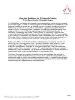
5 Scanning guidelines. ultrasonography . 2017. Apr;36(2):111-119. ultrasonography 36(2), April 2017 111. Ji Young Hwang Anatomy of the lower extremity arteries on the superficial femoral artery medially and the deep femoral artery CT Angiography laterally [10]. The superficial femoral artery descends without prominent branching between the quadratrus and adductor muscle Each lower extremity artery is visible with an accompanying vein, groups in the anteromedial thigh. In the distal thigh, the superficial extending from the iliac artery to the popliteal artery. The anterior femoral artery enters the adductor canal. On leaving the adductor tibial artery, the posterior tibial artery, and the peroneal artery hiatus, the name of the artery becomes the popliteal artery in the are seen with two homonymous veins. The overall anatomy of the popliteal fossa and ends by bifurcating into the anterior tibial artery arteries in the lower extremities is shown on CT angiography in Fig.
6 1. and the tibioperoneal trunk in the posterior aspect of the proximal The common iliac artery splits into the internal iliac artery and the calf [11]. external iliac artery in the pelvic cavity. The external iliac artery is Below the knee, the anterior tibial artery passes from the posterior continuous with the common femoral artery (Fig. 1A). The inguinal to the anterior, and then descends along the interosseous membrane ligament is a landmark for the junction of the external iliac artery behind the anterior tibialis muscle and the extensor muscles in the and common femoral artery. The inguinal ligament is located more anterolateral leg. The tibioperoneal trunk divides into the posterior proximally than the inguinal crease. The common femoral artery is tibial artery medially and the peroneal artery laterally (Fig.)
7 1B). The a short segment, generally about 4 cm long, and bifurcates into posterior tibial artery runs along the intermuscular space between Aorta Knee crease POPA. CIA. EIA ATA Tibioperoneal trunk IIA. CFA Inguinal crease PA PTA. DFA. SFA. ATA PTA. POPA. Knee crease A B. Fig. 1. The anatomy of the lower extremity arteries on computed tomography (CT) angiography. A. On coronal maximal intensity projection (MIP) CT image above the knee, the external iliac artery (EIA) is continuous with the common femoral artery (CFA) which bifurcates into the superficial femoral artery (SFA) and deep femoral artery (DFA). The SFA is continuous with the popliteal artery (POPA). B. On coronal MIP CT image below the knee, the POPA splits anterior tibial artery (ATA) and tibioperoneal trunk which bifurcates into posterior tibial artery (PTA) and peroneal artery (PA).
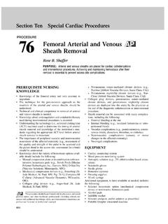
8 CIA, common iliac artery; IIA, internal iliac artery. 112 ultrasonography 36(2), April 2017 Doppler ultrasonography of lower extremity arteries the posterior tibialis muscle and the soleus muscles. The peroneal the transducer, arteries are partially compressed, while veins are artery extends down between the posterior tibialis muscle and the completely collapsed [12]. flexor hallucis longus muscle. Doppler US of the lower extremity begins at the inguinal crease In the ankle and foot region, the anterior tibial artery continues by putting a transducer on the common femoral artery in the into the dorsalis pedis artery distal to the extensor retinaculum [11]. transverse plane with the patient in the supine position (Fig. 2). The The dorsalis pedis artery forms the arcuate artery at the metatarsal common femoral artery is seen lateral to the femoral vein, which base and gives rise to the dorsal metatarsal artery.
9 The posterior is drained from the greater saphenous vein anteromedially at the tibial artery passes behind the medial malleolus of the tibia and inguinal area (Fig. 3A). Just below the inguinal crease, the superficial bifurcates, forming the medial and lateral plantar arteries . The deep femoral artery and the deep femoral artery are present alongside plantar arch from the medial and lateral plantar arteries gives rise to the femoral vein, showing a shape reminiscent of Mickey Mouse's the plantar metatarsal and digital arteries of the foot [11]. face on a transverse scan (Fig. 3B). The common femoral artery, the bifurcated superficial femoral artery and deep femoral artery are US Anatomy of the lower extremity arteries seen in a fallen-Y configuration in a longitudinal scan (Fig. 2). From the proximal to distal thigh, scanning is performed by moving a arteries can be differentiated from veins on US by several transducer distally along the superficial femoral artery deep to the characteristics.
10 First, arteries are round in transverse images, while sartorius muscle. The superficial femoral artery goes together with veins are somewhat oval. Second, arteries are smaller than veins. the femoral vein (Fig. 2). Third, arteries have visible walls and sometimes have calcified The popliteal artery is evaluated from the knee crease level in plaques on the walls. Fourth, when the vessels are compressed by the transverse plane and then traced proximally up to the adductor Supine position Prone position 1 3. 1 GSV CFA. 2. SFA. 3 FV CFA. 2 DF FV. GSV SFA A. 4 DFA. FV. 4. SFA. 5 FV 5. 6. 5 SSV 6. POPV. POPV. POPA POPA. Fig. 2. The steps of color Doppler ultrasonography (US) for the lower extremities above the knee, with the patient's position indicated. The red rectangular boxes are the essential scanning sites and planes for the femoral arteries and the popliteal artery.