Transcription of Doses from Medical X-ray Procedures
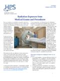
1 Doses from Medical X Ray Procedures Standardized radiation dose estimates can be given for a number of typical diagnostic Medical Procedures . We are not able to give radiation dose estimates for Procedures involving radiation therapy; these need to be handled very carefully on a case by case basis. The tables below give dose estimates for typical diagnostic radiology exams for adults. Doses will change depending on a number of variables including the specific machine and manufacturer, study techniques (the settings of the machine used to produce the radiation in radiology and, in nuclear medicine, the amount of activity administered and the patient s metabolism), and other issues. For comparison, all of us receive about 300 mrem (3 mSv) of radiation exposure to natural background radiation every year. A Medical imaging exam that involves exposure to ionizing radiation must be justified on the basis of benefit to the patient.
2 No practice involving exposure to ionizing radiation should occur unless it produces sufficient benefit to the exposed individual (ICRP 1991). To assist in determining what appropriate practice is, the American College of Radiology has developed Practice Guidelines and Appropriateness Criteria. Practice guidelines are an educational tool designed to assist practitioners in providing appropriate care for patients. Appropriateness criteria are evidence based guidelines to assist referring physicians and other providers in making the most appropriate imaging or treatment decision. The use of Medical exams that would expose children to ionizing radiation needs special consideration. Children have a 3 5 times larger radiation induced cancer mortality risk than adults (ICRP 1991). Most organizations adjust the parameter settings on an x ray machine or the amount of radioactivity administered (for nuclear medicine scans) when performing exams on children; however, it is important to affirm that reduced factors for children s exams have been adopted by the facility in question.
3 The American College of Radiology has developed pediatric CT protocol guidance to assist facilities working to reduce the Doses delivered. Estimates of the dose an individual might receive from one x ray. Single Radiograph Effective dose , mrem (mSv) Skull (PA or AP)1 3 ( ) Skull (lateral)1 1 ( ) Chest (PA)1 2 ( ) Chest (lateral)1 4 ( ) Chest (PA and lateral)2 6 ( ) Thoracic spine (AP)1 40 ( ) Thoracic spine (lateral)1 30 ( ) Lumbar spine (AP)1 70 ( ) Lumbar spine (lateral)1 30 ( ) Abdomen (AP)1 70 ( ) Abdomen3 53 ( ) Pelvis (AP)1 70 ( ) Pelvis or hips3 83 ( ) Bitewing dental film3 ( ) Limbs and joints3 6 ( ) Estimates of the dose an individual might receive if undergoing an entire procedure ( , a lumbar spine series typically consists of five films). Complete Exams Effective dose , mrem (mSv) Intravenous pyelogram (kidneys, 6 films)1 250 ( ) Barium swallow (24 images, 106 sec fluoroscopy)1 150 ( ) Barium meal (11 images, 121 sec fluoroscopy)1 300 ( ) Barium follow up (4 images, 78 sec fluoroscopy)1 300 ( ) Barium enema (10 images, 137 sec fluoroscopy)1 700 ( ) CT head1 200 ( ) CT chest1 800 ( ) CT abdomen1 1,000 (10) CT pelvis1 1,000 (10) CT (head or chest)2 1,110 ( ) PTCA (heart study)3 750 5,700 ( 57) Coronary angiogram3 460 1,580 ( ) Mammogram3 13 ( ) Lumbar spine series3 180 ( ) Thoracic spine series3 140 ( ) Cervical spine series3 27 ( ) References for the two tables 1.
4 Wall BF, Hart D. Revised radiation Doses for typical x ray examinations. The British Journal of Radiology 70:437 439; 1997 (5,000 patient dose measurements from 375 hospitals). 2. National Council on Radiation Protection and Measurements. Sources and magnitude of occupational and public exposures from nuclear medicine Procedures . Bethesda, MD: National Council on Radiation Protection and Measurements; NCRP Report 124; 1996. 3. United Nations Scientific Committee on the Effects of Atomic Radiation. Sources and effects of ionizing radiation, Vol. 1: Sources. New York, NY: United Nations Publishing; 2000. Other references and resources American College of Radiology white paper on radiation dose in medicine: American College of Radiology practice guideline for diagnostic reference levels in Medical x ray imaging: diagnostic International Commission on Radiological Protection. 1990 recommendations of the International Commission on Radiological Protection.
5 Oxford: Pergamon Press; ICRP Publication 60; Ann ICRP 21(1 3); 1991. Glossary background radiation: Background radiation includes radiation from cosmic sources, naturally occurring radioactive materials (including radon), and global fallout (from the testing of nuclear explosive devices). The typically quoted average individual exposure from background radiation is rem per year. becquerel: The becquerel (Bq) is the unit in the International System of Units to replace the curie (see curie). curie: The curie (Ci) is the original term used to describe the amount of radioactive material present or strength of the source. It is based upon the radioactive decay rate of the radionuclide. One curie is equal to x 1010 disintegrations (37 trillion decays) per second (dps); one becquerel is equal to 1 dps. The most common activity levels used in laboratories are the millicurie (mCi) and microcurie ( Ci).
6 A millicurie (mCi) is 1/1,000th of a curie and a microcurie ( Ci) is 1/1,000,000th of a curie. In the International System of Units, the becquerel (Bq) describes the amount of radioactive material present. One curie is equal to x 109 Bq. diagnostic: In medicine, diagnosis or diagnostics is the process of identifying a Medical condition or disease by its signs and symptoms and from the results of various Procedures . As used when referring to Medical exams involving radiation, it is the use of x rays or radioactive materials to identify the Medical condition. dose : dose is a general term used to express (quantify) how much radiation exposure something (a person or other material) has received. The exposure can subsequently be expressed in terms of the absorbed, equivalent, committed, and/or effective dose based on the amount of energy absorbed and in what tissues. effective dose : Radiation exposures to the human body, whether from external or internal sources, can involve all or a portion of the body.
7 The health effects of one unit of dose to the entire body are more harmful than the same dose to only a portion of the body, , the hand or the foot. To enable radiation protection specialists to express partial body exposures (and the accompanying Doses ) to portions of the body in terms of an equal dose to the whole body, the concept of effective dose was developed. Effective dose , then, is the dose to the whole body that carries with it the same risk as a higher dose to a portion of the body. As an example, 8 rem (80 mSv) to the lungs is roughly the same potential detriment as 1 rem (10 mSv) to the whole body based on this idea. exposure: Exposure is commonly used to refer to being around a radiation source; , if you have a chest x ray, you are exposed to radiation. By definition, exposure is a measure of the amount of ionizations produced in air by photon radiation. exposure rate: Exposure rate is the amount of exposure or dose you are receiving per unit time ( , 1 mrem/hour).
8 Gamma rays: Gamma rays are high energy electromagnetic radiation (photons) emitted in an attempt by the radionuclide to become stable, , radioactive decay. Gamma rays have moderate to high penetrating power, are often able to penetrate deep into the body, and generally require some form of shielding, such as lead or concrete. Visible light is also in the form of photons. Gamma photons behave similarly to light, but they are invisible. high level radiation: High level radiation refers to radiation Doses greater than 10 rem to a human body. low level radiation: Low level radiation refers to radiation Doses less than 10 rem to a human body. observable health effect: An observable health effect is a change in physical health that can be detected medically. Observable health effects may include changes in blood cell counts, skin reddening, cataracts, etc. Whether or not it is an observable harmful health effect depends on whether damage to the body has occurred and whether that damage impairs how the body is able to function.
9 Radiation: Radiation is a term commonly used to describe ionizing radiation ( , x and gamma rays, alpha and beta particles, neutrons). Ionizing radiation is radiation that is capable of producing ions by passing through matter. rem: Rem is the term used to describe equivalent or effective radiation dose . In the International System of Units, the sievert (Sv) describes equivalent or effective radiation dose . One sievert is equal to 100 rem. One mrem is one thousandth of a rem. risk: Risk is defined in most health related fields as the probability or odds of incurring injury, disease, or death. roentgen: The roentgen (R) is the term used to describe radiation exposure. This term for exposure only describes the amount of ionization in air. In the International System of Units, the coulomb/kilogram (C/kg) describes radiation exposure. One roentgen is equal to x 10 4 C/kg. safe: Safe, as it is being used in the information on this Web site, is defined as an activity that is generally considered acceptable to us.
10 This is not to say there is absolutely no risk with an activity that is considered safe; there may be a risk from the activity or the exposure to radiation, but it is the same or lower than the risks from everyday actions. At a level of radiation that is considered safe, an effect is either nonexistent or too small to observe. sievert: Sievert (Sv) is the unit in the International System of Units to replace the rem (see rem). therapy: Therapy is the Medical treatment of disease or disorders. With respect to radiation therapy, therapeutic Doses ( , external beam treatments for tumors, radioiodine treatment for thyroid disorders) are significantly greater than those received from diagnostic Procedures ( , chest x rays, CT scans, nuclear medicine Procedures , etc.). x rays: X rays are electromagnetic radiation (photons) that can be emitted from radionuclides or from certain types of devices. Generally, x rays have lower energies than gamma rays, but like gamma rays, x rays can penetrate into the body.