Transcription of Edema: Diagnosis and Management - AAFP Home
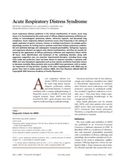
1 102 American Family Physician Volume 88, Number 2 July 15, 2013 Edema: Diagnosis and ManagementKATHRYN P. TRAYES, MD, and JAMES S. STUDDIFORD, MD, Thomas Jefferson University Hospital, Philadelphia, PennsylvaniaSARAH PICKLE, MD, Rutgers Robert Wood Johnson Medical School, New Brunswick, New JerseyAMBER S. TULLY, MD, Cleveland Clinic, Cleveland, Ohio Edema is an accumulation of fluid in the intercellular tissue that results from an abnormal expansion in interstitial fluid volume. The fluid between the interstitial and intravascular spaces is regulated by the capillary hydro-static pressure gradient and the oncotic pressure gradient across the The accumulation of fluid occurs when local or systemic conditions disrupt this equilib-rium (Ta b l e 11-13), leading to increased capil-lary hydrostatic pressure, increased plasma volume, decreased plasma oncotic pressure (hypoalbuminemia), increased capillary permeability, or lymphatic of EdemaHISTORYThe history should include the timing of the edema, whether it changes with position, and if it is unilateral or bilateral, as well as a medication history and an assessment for systemic diseases (Ta b l e 2).
2 Acute swelling of a limb over a period of less than 72 hours is more characteristic of deep venous thrombo-sis (DVT), cellulitis, ruptured popliteal cyst, acute compartment syndrome from trauma, or recent initiation of calcium channel block-ers (Figures 1 and 2). The chronic accumula-tion of more generalized edema is due to the onset or exacerbation of chronic systemic conditions, such as congestive heart failure (CHF), renal disease, or hepatic ,5 Dependent edema caused by venous insuf-ficiency is more likely to improve with eleva-tion and worsen with ,14 Edema associated with decreased plasma oncotic pressure ( , malabsorption, liver failure, nephrotic syndrome) does not change with swelling from compression or compromise of venous or lymphatic Edema is an accumulation of fluid in the interstitial space that occurs as the capillary filtration exceeds the limits of lymphatic drainage, producing noticeable clinical signs and symptoms.
3 The rapid development of generalized pitting edema associated with systemic disease requires timely Diagnosis and Management . The chronic accumulation of edema in one or both lower extremities often indicates venous insufficiency, especially in the presence of dependent edema and hemosiderin deposition. Skin care is crucial in preventing skin breakdown and venous ulcers. Eczematous (stasis) dermatitis can be managed with emollients and topical steroid creams. Patients who have had deep venous thrombosis should wear compres-sion stockings to prevent postthrombotic syndrome. If clinical suspi-cion for deep venous thrombosis remains high after negative results are noted on duplex ultrasonography, further investigation may include magnetic resonance venography to rule out pelvic or thigh proximal venous thrombosis or compression.
4 Obstructive sleep apnea may cause bilateral leg edema even in the absence of pulmonary hyperten-sion. Brawny, nonpitting skin with edema characterizes lymphedema, which can present in one or both lower extremities. Possible secondary causes of lymphedema include tumor, trauma, previous pelvic surgery, inguinal lymphadenectomy, and previous radiation therapy. Use of pneumatic compression devices or compression stockings may be help-ful in these cases. (Am Fam Physician. 2013 ; 88 (2) :102-110. Copy right 2013 American Academy of Family Physicians.) Patient information: A handout on this topic is available at online at This clinical content conforms to AAFP criteria for continuing medical education (CME). See CME Quiz on page disclosure: No rel-evant financial BY CRAIG ZUCKERMAND ownloaded from the American Family Physician website at Copyright 2013 American Academy of Family Physicians.
5 For the private, non-commercial use of one individual user of the website. All other rights reserved. Contact for copyright questions and/or permission 15, 2013 Volume 88, Number 2 American Family Physician 103drainage can result from DVT, venous insufficiency, venous obstruction by tumor ( , tumor obstruction of the iliac vein), lymphatic obstruction ( , from a pelvic tumor or lymphoma), or lymphatic destruction ( , congenital vs. secondary from a tumor, radiation, or filariasis). Bilateral or generalized swelling suggests a systemic cause, such as CHF (especially right-sided), pulmonary hypertension, chronic renal or hepatic dis-ease (causing hypoalbuminemia), protein-losing enter-opathies, or severe ,4,5 Edema can be an adverse effect of certain medications (Ta b l e 31-5).
6 The mechanism often includes the retention of salt and water with increased capillary hydrostatic pressure. Diuretic use may cause volume depletion and reflex stimu-lation of the renin-angiotensin history should also include ques-tions about cardiac, renal, thyroid, or hepatic disease. Graves disease can lead to pretibial myxedema, whereas hypo-thyroidism can cause generalized myx-edema. Although considered a Diagnosis of exclusion, obstructive sleep apnea has been shown to cause edema. One study evaluated the apnea-hypopnea index in patients with obstructive sleep apnea and found that even when adjusted for age, body mass index, and the presence of hypertension and diabetes mellitus, the index was higher in patients who had EXAMINATIONThe physical examination should assess for systemic causes of edema, such as heart failure ( , jugular venous distention, crackles), renal disease ( , proteinuria, oliguria), hepatic disease ( , jaundice, ascites, asterixis), or thyroid disease ( , exophthalmos, tremor, weight loss).
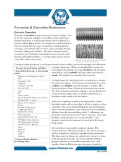
7 Edema should also be evaluated for pit-ting, tenderness, and skin changes. pitting describes an indentation that remains in the edematous area after pres-sure is applied (Figure 3). This occurs when fluid in the interstitial space has a low concentration of protein, which is associated with decreased plasma oncotic pressure and disorders caused by increased capillary pressure ( , DVT, CHF, iliac vein compression).4,16 The phy-sician should describe the location, tim-ing, and extent of the pitting to determine treatment response. Lower extremity examination should focus on the medial malleolus, the bony portion of the tibia, and the dorsum of the foot. pitting edema Table 1. Systemic and Localized Causes of EdemaCauseMechanism of actionSystemicAllergic reaction, urticaria, and angioedemaIncreased capillary permeabilityCardiac diseaseIncreased capillary permeability from systemic venous hypertension; increased plasma volumeHepatic diseaseIncreased capillary permeability from systemic venous hypertension; decreased plasma oncotic pressure from reduced protein synthesisMalabsorption/protein-calorie malnutritionReduced protein synthesis leading to decreased plasma oncotic pressureObstructive sleep apneaPulmonary hypertension resulting in increased capillary hydrostatic pressurePregnancy and premenstrual edemaIncreased plasma volumeRenal diseaseIncreased plasma volume.
8 Decreased plasma oncotic pressure from protein lossLocalizedCellulitisIncreased capillary permeabilityChronic venous insufficiencyIncreased capillary permeability caused by local venous hypertensionCompartment syndromeIncreased capillary permeability caused by local venous hypertensionComplex regional pain syndrome type 1 (reflex sympathetic dystrophy)Neurogenically mediated increased capillary permeabilityDeep venous thrombosisIncreased capillary permeabilityIliac vein obstructionIncreased capillary permeability caused by local venous hypertensionLipedemaAccumulation of fluid in adipose tissueLymphedemaPrimary: congenital lymphedema, lymphedema praecox, lymphedema tardaSecondary: from axillary lymph node dissection, surgery ( , coronary artery bypass graft, inguinal lymphadenectomy), trauma, radiation, tumor, filariasisLymphatic obstructionMay-Thurner syndrome (compression of left iliac vein by right iliac artery)Increased capillary permeability caused by local venous hypertension from compressionInformation from references 1 through 2.
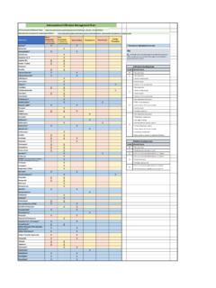
9 Diagnosis and Management of Common Causes of Localized EdemaEtiologyOnset and locationExamination findingsEvaluation methodsTreatmentUnilateral predominance Chronic venous insufficiencyOnset: chronic; begins in middle to older ageLocation: lower extremities; bilateral distribution in later stagesSoft, pitting edema with reddish-hued skin; predilection for medial ankle/calfAssociated findings: venous ulcerations over medial malleolus; weeping erosionsDuplex ultrasonographyAnkle-brachial index to evaluate for arterial insufficiencyCompression stockingsPneumatic compression device if stockings are contraindicated Horse chestnut seed extractSkin care ( , emollients, topical steroids) Complex regional pain syndrome type 1 (reflex sympathetic dystrophy)Onset: chronic; following trauma or other inciting eventLocation: upper or lower extremities; contralateral limb at risk regardless of traumaSoft tissue edema distal to affected limbAssociated findings: (early) warm, tender skin with diaphoresis; (late) thin, shiny skin with atrophic changesHistory and examinationRadiographyThree-phase bone scintigraphyMagnetic resonance imagingSystemic steroidsTopical dimethyl sulfoxide solution Physical therapyTricyclic antidepressantsCalcium channel blockersDVTO nset: acuteLocation: upper or lower extremitiesPitting edema with tenderness, with or without erythema.
10 Positive Homans signD-dimer assayDuplex ultrasonographyMagnetic resonance venography to rule out pelvic or thigh DVT (if clinical suspicion is high), or extrinsic venous compression (May-Thurner syndrome in patients with unexplained left-sided DVT)Consider hypercoagulability workupAnticoagulation therapyCompression stockings to prevent postthrombotic syndromeThrombolysis in select patientsLymphedemaOnset: chronic; insidious; often following lymphatic obstruction from trauma or surgeryLocation: upper or lower extremities; bilateral in 30% of patientsEarly: dough-like skin; pittingLate: thickened, verrucous, fibrotic, hyperkeratotic skinAssociated findings: inability to tent skin over second digit, swelling of dorsum of foot with squared off digits, painless heaviness in extremityClinical diagnosisLymphoscintigraphyT1-weighted magnetic resonance lymphangiographyComplex decongestive physiotherapyCompression stockings with adjuvant pneumatic compression devicesSkin careSurgery in limited casesBilateral predominance LipedemaOnset: chronic; begins around or after pubertyLocation: predominantly lower extremities; involves thighs, legs, buttocks; spares feet, ankles, and upper torsoNonpitting edema; increased distribution of soft, adipose tissueAssociated findings: medial thigh and tibial tenderness.