Transcription of EDUCATIONAL COMMENTARY PERIPHERAL BLOOD …
1 EDUCATIONAL COMMENTARY PERIPHERAL BLOOD FINDINGS IN A CASE OF HAIRY CELL LEUKEMIA American Proficiency Institute 2018 1st Test Event 1 EDUCATIONAL COMMENTARY is provided through our affiliation with the American Society for Clinical Pathology (ASCP). To obtain FREE CME/CMLE credits, click on Earn CE Credits under Continuing Education on the left side of the screen. Learning Outcomes On completion of this exercise, the participant should be able to discuss morphologic characteristics of normal PERIPHERAL BLOOD leukocytes; identify distinguishing morphologic features of hairy cells, normal lymphocytes, and reactive lymphocytes; and describe the cytochemical characteristic and immunophenotyping results in hairy cell leukemia.
2 Case History A CBC with differential was ordered on a 71 year old male patient with weakness, fatigue, and weight loss. His CBC results are as follows: WBC= x 109/L, RBC= x 1012/L, Hgb= g/dL, Hct=32%, MCV= fL, MCH= pg, MCHC= g/dL, Platelet=94 x 109/L, RDW-CV= %. Introduction The patient is a 71 year old man diagnosed with hairy cell leukemia, a malignant lymphoproliferative condition. This disorder frequently causes weakness, fatigue, and weight loss, symptoms experienced by this patient. The patient s complete BLOOD cell count values indicate anemia, with low hemoglobin and hematocrit results explaining his weakness and fatigue; he also has thrombocytopenia.
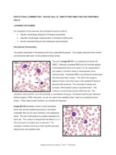
3 Additional symptoms reported in hairy cell leukemia can include infection and splenomegaly. The images provided for initial identification and discussion represent examples of both normal PERIPHERAL white BLOOD cells and hairy cell lymphocytes. COMMENTARY Image BCI-01 is a classic hairy cell. Hairy cells vary in size but often are larger than normal lymphocytes. The cell is generally round or oval; the nuclei are also usually round or oval, but may be indented. Hairy cell chromatin is often finer than that seen in normal lymphocytes, although this feature is not evident in this particular cell. Sometimes nucleoli are visible, but they are not seen in this example.
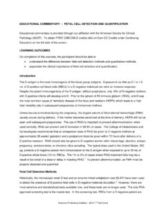
4 Hairy cells derive their name from their distinctive frayed cytoplasmic margins. These projections are EDUCATIONAL COMMENTARY PERIPHERAL BLOOD FINDINGS IN A CASE OF HAIRY CELL LEUKEMIA (cont.) American Proficiency Institute 2018 1st Test Event 2 often irregular, blunted, and fine and appear hairlike. The cytoplasm is generally a pale or grayish blue and moderate to abundant. Image BCI-02 is an eosinophil. Eosinophils are about the same size as segmented neutrophils. The nuclei are frequently bilobed, although it is difficult to see how many lobes are present in this cell. The nuclear chromatin is condensed and clumped.
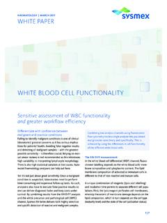
5 Eosinophils are distinguished by their numerous large, red-orange cytoplasmic granules. These granules have been referred to as copper pennies and give the cytoplasm an overall pinkish color. Image BCI-03 is a neutrophil. Note that this cell is similar in size to the eosinophil discussed in Image BCI-02. Segmented neutrophils have two to five nuclear lobes connected by thin strands of chromatin. The chromatin is condensed and clumped, as seen in the eosinophil. However, in contrast to the eosinophil, the cytoplasm in the neutrophil has numerous granules that are pink or pink-violet. The cytoplasm in a neutrophil appears pinkish because of these cytoplasmic granules.
6 A lymphocyte is identified in Image BCI-04. This is an example of a small, resting lymphocyte, although normal lymphocytes vary in size. The nucleus may be round, as in this cell, or slightly indented. The nuclear chromatin is condensed and clumped. These cells typically have a high nuclear to cytoplasmic ratio, so the blue cytoplasm is often scanty, just rimming the nucleus. Note that larger lymphocytes may have a few azurophilic granules, but small lymphocytes usually have no granules. Image BCI-05 is a monocyte. Monocytes are the largest cells normally present in the PERIPHERAL BLOOD . The nuclei in monocytes may appear round, oval, lobulated, or kidney shaped.
7 The nuclear chromatin is minimally clumped and stains EDUCATIONAL COMMENTARY PERIPHERAL BLOOD FINDINGS IN A CASE OF HAIRY CELL LEUKEMIA (cont.) American Proficiency Institute 2018 1st Test Event 3 lighter shades of pink or purple; nucleoli should not be visible. The cytoplasm in monocytes is often abundant and blue-gray. Sometimes cytoplasmic extensions may be seen. The cytoplasm stains unevenly and may look rough, bumpy, or as if grains of sand are present. Cytoplasmic vacuoles are frequently present. The cell selected for Image BCI-06 is another hairy cell. This cell is similar to the hairy cell shown in Image BCI-01, but this cell has more numerous cytoplasmic projections, or hairs.
8 It also has nuclear chromatin that is slightly less clumped and finer than the nuclear chromatin seen in Image BCI-01, but which is still characteristic of a hairy cell. The final image in this testing event, Image BCI-07, is a reactive lymphocyte. Reactive lymphocytes are distinguished by a wide variety of morphologic features. These variations in cellular characteristics reflect the heterogeneity of immune responses, as these lymphocytes are, by definition, reacting to an abnormal stimulus, often resulting from a viral infection. It is not unusual to see a small percentage of reactive lymphocytes on a normal PERIPHERAL BLOOD smear.
9 Likewise, it is not unexpected to see a reactive lymphocyte on a BLOOD smear from a patient with hairy cell leukemia. Leukemia causes great stress to the body s immune system. Although reactive lymphocytes demonstrate many different morphologic characteristics, some generalizations can be made. Reactive lymphocytes are usually large cells. Their nuclei vary in shape and may be round, oval, indented, or lobulated. The nuclear chromatin is often more fine and open than that seen in a nonreactive lymphocyte. Areas of parachromatin may be more visible, and nucleoli may be evident and prominent. Cytoplasm is typically abundant in reactive lymphocytes.
10 The color varies and may be gray, pale blue, or deep blue. The cytoplasmic margin is often indented by surrounding red BLOOD cells, with darker blue evident at this cellular interface. Note the darker blue borders where this cell interacts with nearby erythrocytes. Also, note how the cytoplasm is a lighter blue near the nucleus, another general feature of reactive lymphocytes. It is possible to see azurophilic granules and vacuoles in the cytoplasm of reactive lymphocytes. The morphologic variety associated with reactive lymphocytes can be contrasted with the homogeneity of morphologic features seen in a malignant condition such as hairy cell leukemia.