Transcription of ELISA technical guide and protocols
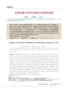
1 TECH TIP # 65 ELISA technical guide and protocols Table of Contents Introduction ..2 Variations between ELISA protocols ..3 A. Antigen B. Direct vs. Indirect Detection of C. Biotin Signal Amplification ..3 D. Fluorescence ..4 E. Enzymatic Detection ..4 F. Substrates ..4 Factors affecting assembly of the immune A. Types of Plate and Coupling Options ..5 B. Antibodies ..6 C. Blocking Buffer ..6 D. Target E. Enzyme Conjugate ..7 F. Washing the Plate ..7 G. Substrates and Signal Detection ..7 Common Occurrences and Explanations ..7 A. Low Signal-to-Noise B. Signal Fades Quickly (generally seen with chemiluminescence only) ..8 C. Substrate Forms a Precipitate or Plate Visibly Glows in the Dark ..8 D. Fluorescent Signal is Low ..8 E. Poor Standard Curve Linearity and Dynamic General Capture/Sandwich ELISA A.
2 Materials Required ..10 B. Preparation of C. Preparation of D. Modifications to the ELISA protocol ..11 A. Performing Direct Antigen B. Using an Enzyme Labeled Secondary Antibody ..11 C. Using an Alkaline Phosphatase D. Using Fluorescence ..11 E. Using F. Using Chemiluminescence ..11 G. Competition ELISAs ..12 ELISA Optimization ..12 A. Optimizing Capture Antibody B. Optimizing the Blocking Buffer ..12 C. Optimizing the Standard Diluent ..12 D. Optimizing Sample Concentration ..13 E. Optimizing the Detection Antibody Concentration ..13 F. Optimizing the Enzyme Conjugate Concentration ..13 G. Optimizing Signal Detection ..13 Appendix ..13 References ..14 Pierce Biotechnology PO Box 117 (815) 968-0747 3747 N. Meridian Road Rockford, lL 61105 USA (815) 968-7316 fax Introduction The enzyme linked immunosorbent assay ( ELISA ) is a powerful method for detecting and quantifying a specific protein in a complex mixture.
3 Originally described by Engvall and Perlmann (1971), the method enables analysis of protein samples immobilized in microplate wells using specific antibodies. The technique has revolutionized immunology and is commonly used in medical research laboratories. ELISA also has commercial applications, including the detection of disease markers and allergens in the diagnostic and food industries. The ELISA method was made possible because of scientific advances in a number of related fields. Technology enabling the production of antigen-specific monoclonal antibodies by Kohler and Milstein (1975) led to their use as probes for detecting individual molecules in complex protein mixtures or tissue samples. Initially, detection was achieved by radioimmunoassay using antibodies labeled with radioisotopes, but because of health risks alternatives were sought.
4 Avramais (1966, 1969) and Pierce (1967) developed methods to chemically link antibodies to biological enzymes whose activities produce a measurable signal with solutions containing appropriate substrates. With the development of fluorescence technology, signal generation using fluorophore-labeled antibodies has also become prevalent, especially in multiplex arrays. Although many variants of ELISA have been developed and used in different situations, they all depend on the same basic elements: 1. Coating/Capture: direct or indirect immobilization of antigens to the surface of polystyrene microplate wells. 2. Plate Blocking: addition of irrelevant protein or other molecule to cover all unsaturated surface-binding sites of the microplate wells. 3. Probing/Detection: incubation with antigen-specific antibodies that affinity-bind to the antigens.
5 4. Signal Measurement: detection of the signal generated via the direct or secondary tag on the specific antibody. In a typical assay designed to detect an antigen in a complex protein mixture, the antigen is immobilized either by direct adsorption or via an antibody adsorbed to the wells of a microplate. The plate is blocked and the antigen is probed with a specific detection antibody. The detection antibody may be directly labeled with a signal-generating enzyme or fluorophore or it may be secondarily probed with an enzyme- or fluor-labeled secondary antibody (or avidin-biotin chemistry, see below). For enzymatic detection, the appropriate enzyme substrate is added. The signal observed is proportional to the amount of antigen in the sample. Washing between steps ensures that only specific (high-affinity) binding events are maintained to cause signal at the final step.
6 Diagram of popular ELISA formats. Pierce Biotechnology PO Box 117 (815) 968-0747 3747 N. Meridian Road Rockford, lL 61105 USA (815) 968-7316 fax 2 Variations between ELISA protocols A. Antigen Immobilization Antigen immobilization varies between two principle techniques. In a traditional (direct coating) ELISA , antigens are directly attached to the plate by passive adsorption, usually using a carbonate/bicarbonate buffer at pH >9. Most but not all proteins bind tightly to the polystyrene surface of microplates in alkaline conditions. However, if antigen is present at low levels or does not adhere well to the plastic, then the alternative sandwich or capture ELISA may be used. In capture (indirect coating) ELISA , antigen-specific antibody is adsorbed onto the plastic, which in turn binds and immobilizes the antigen upon incubation with the antigen sample.
7 Attachment of the antibody is typically achieved using the same carbonate/bicarbonate buffer at pH >9, or in rare instances pre-activated plates are used for a more directed attachment approach. Sandwich ELISAs have become very popular when using complex protein samples because only the specific antigen becomes immobilized rather than the entire sample of proteins. The more antigen that is immobilized, the higher is the potential sensitivity of the assay . Sandwich ELISAs require two different antibodies that bind specifically to the antigen (each reacting with a different epitope). The first antibody (bound to the plate) is called the capture antibody or coating antibody, whereas the second antibody detects the immobilized antigen and is called the detection antibody. Such antibodies are known as matched pairs ; they must be validated to work in combination, as they must not compete for binding to the antigen for accurate results to be possible.
8 Combinations of monoclonal and polyclonal antibodies can be used; it is more common to use the monoclonal as the coating antibody and the polyclonal as the detection antibody. Sandwich ELISAs sometimes require more optimization than traditional ELISAs but usually the signal-to-noise (S:N) ratios are higher. B. Direct vs. Indirect Detection of Antigen Direct detection involves labeling of the detection antibody with an enzyme or an alternative signaling molecule such as a fluorophore. Indirect detection involves an additional probing step using another antibody or streptavidin that is labeled with a detectable tag. This additional probe is called the secondary antibody, as its sole purpose is to deliver the measurable signal tag by binding to the detection (primary) antibody. Direct detection is generally faster than indirect detection and potential background signal from secondary antibody cross-reactivity with the coating antibody is also eliminated.
9 Even in the best of circumstances however, direct detection cannot provide the signal amplification gained from the use of a secondary antibody or avidin/biotin systems. Consequently direct detection is generally less sensitive than indirect detection and is best used only when the target is relatively abundant. C. Biotin Signal Amplification Biotin is a small (MW 244) vitamin molecule that is easily modified so that it can be chemically attached to proteins, antibodies and other biomolecular probes of interest. Avidin and streptavidin are proteins that originate from different sources but have nearly identical functions in binding very strongly and specifically to the biotin molecule. Thermo Scientific NeutrAvidin Protein is a specially modified form of avidin. Hereafter in this document, avidin and streptavidin will be used interchangeably to mean any one of these biotin-binding proteins.
10 The avidin-biotin affinity system is frequently employed in the design of tagging and detecting systems for ELISA . A common adaptation of indirect detection is to amplify the signal using avidin-biotin chemistry. There are two approaches. The first method involves using a biotinylated detection antibody, which is probed using avidin or streptavidin protein conjugated to either horseradish peroxidase (HRP) or alkaline phosphatase (AP) enzymes. The second approach also employs a biotinylated detection antibody, but it is probed with a pre-incubated mixture of avidin and biotinylated enzyme, a process known as avidin-biotin complex (ABC) signal amplification. Diagram of the signal amplification that is possible with the ABC system. Signal amplification occurs through two mechanisms with these approaches.