Transcription of Emergency Neurological Life Support Traumatic …
1 Emergency Neurological life Support Traumatic Brain Injury Version: Last Updated: 19-Mar-2016. Traumatic Brain Injury Table of Contents Emergency Neurological life Support .. 1. Traumatic Brain 1. Checklist .. 3. 3. Analgesia and Sedation .. 4. Avoid Hypoxia and Hypotension .. 5. Keep SBP > 90 mmHg, O2 Sat > 90% .. 5. Coagulopathy .. 6. Recognition and treatment .. 6. Diagnosis .. 7. What constitutes TBI? .. 7. Hemicraniectomy .. 8. And other surgical interventions .. 8. Initial Hospital Management .. 9. If not done 9. Medical Therapy ..10. Evaluation and management in the field ..11. Control and prevention ..12. Treat Elevated ICP ..13. ICP Monitoring- Indications and treatment algorithm ..13. Traumatic Brain Injury Page 3. Checklist Secure Airway SBP > 90 mmHg and O2 saturation > 90%. C-spine precautions Head CT. Treat herniation Neurologic examination Communication Patient age and mechanism of injury Pre-injury health, including home medications Head CT findings Post resuscitation GCS with detailed neurologic exam Completed interventions Focal motor findings Coagulation status and other pertinent laboratory findings Other injuries State of cervical spine - cleared, not cleared, injury Current vital signs Traumatic Brain Injury Page 4.
2 Analgesia and Sedation Patients with severe TBI typically require tracheal intubation for airway protection. Depending on the level of consciousness and agitation, sedation is often required. Concomitant skull and body injuries also require analgesia (rib fractures for example). See the ENLS protocol Airway, Ventilation and Sedation for a discussion on medications and methodology. For extubated patients care must be given to prevent oversedation with respiratory depressant medications (opiates and IV benzodiazepines). Traumatic Brain Injury Page 5. Avoid Hypoxia and Hypotension Keep SBP > 90 mmHg, O2 Sat > 90%. Hypotension (SBP < 90 mmHg in adults; < 70 mmHg + age X 2 for children) in the setting of TBI is harmful. At any point in the initial encounter, treat with IV fluid bolus (500 ml 1 L of crystalloid in adults; 20 ml/kg for children). Avoid use of D5W. Oxygenation should be maintained with supplemental oxygen as needed to keep O2 Sat >. 90%. Avoid hyperoxia. Traumatic Brain Injury Page 6.
3 Coagulopathy Recognition and treatment Indicated if known or suspected coagulopathy: recent elevated PT/INR/PTT. low platelets history or physical examination consistent with end-stage hepatic or renal disease on anticoagulant therapy on antiplatelet therapy Consider the following: Plasma or PCC and vitamin K - for patients on warfarin Consider FFP for patients with liver dysfunction with coagulopathy Platelets - for patients with conditions with low or malfunctioning platelets DDAVP - for patients with end-stage renal disease or on certain anti-platelet agents See ENLS reference Pharmacotherapy for detailed antidotes and dosing. Sidebar common pitfalls In most cases, reversal can begin immediately according to empiric guidelines and does not require laboratory values or confirmation. Reversal of anticoagulation is a complex subject, and in some cases, such as in patients with hemophilia and other bleeding dyscrasias, it may be necessary to obtain specialist consultation from a hematologist.
4 Reversal of antiplatelet agents such as ASA, clopidogrel and ticlopidine is controversial. Evidence for platelet transfusion in this cohort is equivocal. Some authors recommend the use of DDAVP despite lack of conclusive benefit. Traumatic Brain Injury Page 7. Diagnosis What constitutes TBI? Traumatic Brain Injury (TBI): Severe TBI: Mechanism consistent with TBI and/or physical signs of trauma in unconscious patient, with a Glasgow Coma Scale (GCS) < 9. It is important to consider other treatable causes of decreased level of consciousness. Every attempt should be made to identify and reverse vascular, metabolic, infectious, environmental, toxicological and other non- Traumatic causes. These causes may co-exist with TBI. The GCS should be obtained through interaction with the patient ( by giving verbal commands or if those unable to respond by applying a painful stimulus). The GCS should be assessed after appropriate resuscitation and before the administration of sedative or neuromuscular blocking agents Diagnosis of TBI - recognition of TBI depends on consideration of: Physiology ( GCS).
5 Anatomy (scalp laceration, depressed skull fracture). Mechanism of injury ( fall > 20 feet, MVA > 30 mph). Concussion Concussion is recognized as a clinical syndrome of biomechanically induced alteration of brain function, typically affecting memory and orientation, which may involve loss of consciousness (LOC). This protocol will not address concussion further Topic Co-Chairs: Rachel Garvin, MD. Chitra Venkatasubramanian, MD. Angela Lumba-Brown, MD. Chad M. Miller, MD. Traumatic Brain Injury Page 8. Hemicraniectomy And other surgical interventions Surgical removal of hematoma (subdural, epidural, intracerebral) depend on the patient's clinical status and the judgment of treating physicians. Some general guidelines include Epidural hematoma or subdural hematomas > 1 cm thick and midline shift > 5 mm Intracerebral hemorrhage > 50 cc in volume or > 3 cm in diameter Penetrating skull injury Depressed skull fracture Refractory ICP. Decompressive hemicraniectomy, either unilateral or bilateral, in select patients can markedly lower ICP and likely improve outcomes; proper patient selection is still being elucidated.
6 Traumatic Brain Injury Page 9. Initial Hospital Management If not done prehospital Spinal precautions to be maintained at all times Advanced airway management to ensure: a) airway protection to maintain oxygen saturation > 90%, b) control of ventilation (if inadequate or inappropriate). See ENLS. protocol Airway, Ventilation and Sedation. Obtain CXR. Continuous monitoring of oxygenation, blood pressure, cardiac rhythm and PCO2. Obtain parenteral access (IV or IO). Diagnose hypoglycemia: if hypoglycemic give D50% 50 ml IV. Obtain CT Head without contrast Consider spine imaging; see ENLS protocol Traumatic Spine Injury. Neurosurgical consultation may be necessary depending on the severity of the injury and the patient's clinical status. Findings that should prompt neurosurgical consultation include: GCS < 13. The patient has seizures Lateralizing findings on Neurological examination, including unequal pupils or focal weakness Abnormal head CT scan Head CT is not consistent with clinical signs CSF leak, or signs of basal skull fracture Penetrating skull injury Cerebrovascular injury Suspected cervical spine injury Sidebar common pitfalls Although a Glasgow Coma Scale of 8 or less during the initial evaluation is an indication for endotracheal intubation; severe extracranial injuries or a rapidly declining mental status may also be indications.
7 Patient can be ventilated with 100% O2 until ABG values are available. Any adjustments should maintain SaO2 > 90%. Traumatic Brain Injury Page 10. Medical Therapy Initial treatment may include: Positioning: ensuring head midline and at 30 degrees HOB elevation if able Analgesic - Fentanyl preferred, ketamine is an option Sedation - Propofol or precedex, depending on patient's vital signs (benzodiazepines are not recommended in TBI patients). Both agents likely drop MAP which will decrease CPP (MAP-ICP). Transfuse RBCs if active bleeding or hemoglobin < 7 gm/dl Use pressors if needed to maintain Goal CPP of 50-70 mmHg Neuromuscular blockers only if shivering or difficulty ventilating patient Control body temperature; avoid fever. Consider temperature management protocol Mannitol or hypertonic saline: Administer 20% mannitol g/kg IV as a rapid (5. minutes) IV infusion; If BP (systolic) < 90 mmHg in adults, hypertonic saline rather than mannitol should be used - administer 3% NaCl 250ml IV over minutes Maintain normocarbia or mild hypocarbia.
8 PbTO2 or SjvO2 monitoring may be beneficial in avoidance of injury due to hyperventilatory ischemia. If needed for ICP. control, hyperventilate to target a PCO2 of 28-35 mmHg (20 breaths a minute in adult) until other urgent, definitive strategies can be employed (typically surgical). Vasopressors may be needed to maintain cerebral perfusion during the process of volume resuscitation If ICP is refractory to medical therapy, hemicraniectomy or lesion removal should be considered. Sidebar common pitfalls Hypotension (systolic BP< 90 mmHg) should prompt rapid discontinuation of mannitol. To administer hypertonic saline, serum sodium should be < 160 mEq/L. Traumatic Brain Injury Page 11. Prehospital Evaluation and management in the field Spinal precautions to be maintained at all time Basic and advanced airway management as indicated to maintain oxygen saturation greater than 90%. See ENLS protocol Airway, Ventilation and Sedation. Normal breathing should be maintained (EtCO2 35-40 mmHg) and hyperventilation avoided (EtCO2 < 35 mmHg) unless there are signs of herniation (see below).
9 When hyperventilation is indicated, 20 breaths per minute in the adult can be used as temporary measure until signs of herniation resolve Continuous monitoring of oxygenation (pulse oximetry) and blood pressure In the adult, systolic BP should be > 90 mmHg Hypotensive patients should be treated with isotonic or hypertonic fluids Obtain IV access Diagnose hypoglycemia: if hypoglycemic give D50% 50 ml IV. Assess Glasgow Coma Score and pupils Common pitfalls The use of neuromuscular blocking medications to facilitate intubation (rapid sequence intubation) in the field worsened outcomes in one large study. If it is performed for other indications, monitoring of oxygenation, blood pressure and end- tidal CO2 should take place. Hypo- and hyperventilation should both be avoided. If EtCO2 measurement is available, this should be in the range of 35 to 40 mmHg. Hyperventilation to decrease PCO2 to between 28-35 mm Hg is only indicated for patients with signs of herniation (rapidly decreasing LOC, particularly with changes in pupil reactivity).
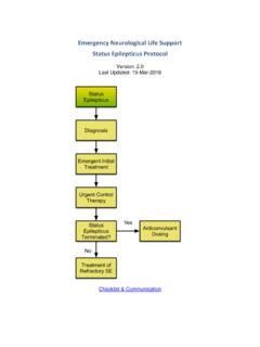
10 Intravenous fluid boluses (500cc to 1L of crystalloid or smaller volumes of hypertonic saline solutions) may be given in adult trauma victims with systolic BP < 90 mmHg or with signs of hypoperfusion ( poor capillary refill). Pupils should be measured after resuscitation and evidence of orbital trauma noted Pupil asymmetry is defined as > 1mm difference in diameter A fixed pupil is defined as < 1 mm decrease in size in response to bright light Signs of herniation include: dilated and nonreactive pupils, asymmetric pupils, motor exam that demonstrates extensor posturing or no response or progressive decline in neurologic condition (decrease in GCS > 2 points). Traumatic Brain Injury Page 12. Seizures Control and prevention If seizure activity was witnessed, or the patient has a depressed level of consciousness, or the head CT is abnormal, it is recommended to treat with anti-epileptic drugs unless there is a known allergy. Phenytoin 20 mg/kg IV no faster than 50 mg/minute Levitiracetam 20 mg/kg is also an option For patients who develop status epilepticus, refer to the ENLS protocol Status Epilepticus.