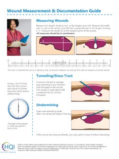
Transcription of Examination Of The Cardiovascular System
1 Examination Of The Cardiovascular SystemPOM -October 9, 2019 Charlie Goldberg, Exam Includes appropriate history and ROS Vital Signs: Blood pressure; Pulse: rate, rhythm, volume Assessment distal vasculature (legs, feet, carotids) vascular disease (atherosclerosis) is a systemic illness ! Pulmonary Exam (coming soon) 4basic PE components: Observation, Palpation, Percussion (omitted in cardiac exam)& AuscultationThoughts On Gown Management & Appropriately/Respectfully Touching Your Patients Several Sources of Tension: Area examined reasonably exposed yet patientmodesty preserved Palpate sensitive areasto perform accurate exam -requires touching people w/whomyou ve little acquaintance awkward, particularly if opposite gender Exam not natural/normalpart of interpersonal interactions-as newcomers to medicine, you re particularly aware & hence sensitive a good thing!
2 Keys To Performing a Respectful & Effective Exam Explain what you re doing (& why) before doing it acknowledge elephant in the room ! Expose minimum amount of skin necessary - artful use of gown & drapes (males & females) Examining heart & lungs of female patients: Ask patient to remove bra prior and/or learn to work around bra Expose side of chest to extent needed Enlist patient s assistance positioning breasts to enable cardiac exam Don t rush, act in a callous fashion, or cause pain don t examine body parts thru gown: Poor technique You ll miss things You ll lose points on scored exams (OSCE, CPX, USMLE)!Observation Pay attention to: Chestshape Shortness of breath(@ restor walking)?
3 Sittingupright? Able to speak? ? Visible impulseon chest wall from vigorously contracting ventricle (rare)Surface AnatomyFinding The Sternal Manubrium Junction (aka Angle of Louis) Key To Identifying Valve AreasManubriumSternumSternalManubriumJun ction(Angle ofLouis)ClavicleClavicle1stRib2ndRibSter numManubriumSternalManubriumJunction2ndI CSM anubriumslopes in one direction while Sternumangles in different direction. Highlighted by q-tips intersection defines Sternal Manubrium JunctionValves And Surface Anatomy Areas of auscultation correlate w/rough location of each valve Where you listen will determine what you hear!More Anatomy Group: -TechniqueLeft ventricle-Fingersacross chest, under breast (explain 1st) Point of Maximal Impulse (PMI) apex ventricle that pin-points w/finger tip.
4 ~70% of patients -if not palpable, repeat w/patient on L side-Sizeof LV increased dimension if PMI shifted to L of mid-clavicular line Vigorof contraction Palpable thrill(rare) -associated w/regurgitant or stenotic murmurs (feels like sensation when kink garden hose)For Female PatientsFor Male PatientsPalpation Technique (cont) Right ventricle: Vigor of contractility heelof R hand along sternum Rarely abnormal with RV (pulmonary hypertension)Auscultation: Using Your StethescopeThey all work -most important part is what goes between the ear pieces!Diaphragm Higher pitched soundsBell Lower pitchedWhat Are We Listening For? Normal valveclosurecreates sound FirstHeart Sound =s S1 closure of Mitral,Tricuspidvalves SecondHeart Sound =s S2 closure of Pulmonic, AorticvalvesCourtesy Wilbur Lew, animation linking cardiac events to Wiggers, ECG and Heart Sounds: Are We Listening For?
5 (cont) Systole=s time between S1 Diastole=s time between S2& S1 Normally, S1& S2 =distinct sounds Physiologic splitting=s 2 components of second heart sound (Aortic& Pulmonicvalve closure) audible w/inspirationBlaufussSimulated Physiologic Splitting of S2: #Auscultation Technique Patient lying@ 30-45degree incline Chest exposed(male) or loosely fitted gown (female) need to see area where placing stethoscope stethoscope must contact skin Stethoscope w/diaphragm(higher pitched sounds) engagedNO!NO!QuickTime and a decompressorare needed to see this !NO!Remember Don t Examine Thru Clothing or Snake Stethoscope Down Shirts/Gowns !Exam Options When Listening toFemale PatientsAuscultation Technique (cont) over Aortic area 2ndRightIntercostal Space (ICS) Use Angle of Louis as (2ndL ICS) down sternal border Tricuspidarea (4thL ICS) towards Mitral area (4thICS, mid-clavicular) Listen in ~ 6 places-precise total doesn t matter gives you sense of change In sounds as change locationAuscultation In each area, ask yourself: Do I hear S1?
6 Do I hear S2? Which is louder& what are relative intensities? Interval between S1& S2 (systole) is shorterthen between S2& S1(diastole) Can also determine timingby simultaneously feelingpulse (a systolic event) Listenfor physiologic splittingof 2nd heart sound w/inspirationMurmurs Murmurs: Sound created by turbulent flowacross valves: Leakage(regurgitation) when valve closed Obstruction (stenosis) toflow when normally open SystolicMurmurs: Aortic stenosis, Mitral regurgitation(Pulmonary stenosis, Tricuspid regurgitation) DiastolicMurmurs: Aortic regurgitation, Mitral stenosis(Pulmonary regurgitation, Tricuspid stenosis) Murmurs (cont) Characterized by: position in cycle, quality, intensity, location, radiation.
7 Can try to draw it s shape: Intensity Scale:1 barely audible 2-readily audible 3-even louder 4-loud + thrill 5-audible with only part of diaphragm on chest 6 audible w/out stethoscope intensity doesn t necessarily correlate w/severity Some murmursbest appreciated in certain positions:Mitral:patient on L side; Aortic:sittingup and leaning forward Example Mitral Regurgitation: Holosystolic, loudest in mitral area, radiates towards : Heart Sound Simulator: Heart Sounds S3 & S4 Ventricularsounds, occur during diastole normal in young patient (~ < 30 yo) usually LV, rarely RV S3 follows S2 caused by blood from LA colliding w/ left over blood in LV associated w/heart failure. S4 precedes S1 caused during atrial systole when blood squeezed into non-compliant LV associated w/HTNE xtra Heart Sound (cont) S3& S4 are soft, low pitched Best heart w/bell, laid over LV, w/patient lying on L side(brings apex of heart closer to chest wall) Abnormal beyond age ~30 When present, S3 or S4are referred to as gallops University of Washington, Simulated S3& S4 An Ordered Approach Do I hear S1?
8 Do I hear S2? Listen in each major valvulararea think about which sound should be loudest in each location (S1 loudest region of TV & MV, S2 loudest AV & PV) Do I hear physiologic splittingof S2? Do I hear something before S1 (an S4) or after S2 (an S3)? Do I hear murmurin systole? In diastole? Ifa murmurpresent, note: intensity, character, duration, radiation As listen, thinkabout mechanical eventsthat generate the Arteries Anatomy Palpation(easide separately!) Rhythm Fullness Auscultation Radiation of murmurs ? Intrinsic atherosclerosis may produce shsshhing noise known as Venous Pressure (JVP) Anatomyof Internal Jugular Vein Straight line with RA Manometer reflecting CentralVenous Pressure (CVP) Technique Findcorrect area helps to first identify SCM & triangle it forms w/clavicle Look for multi-phasicpulsations( a , c & v waves) Isolatefrom carotid pulsations, respirations Tangential lighting Hepatojugular reflux(gentle pressure over liver pushes blood back into IJ & makes pulsations more apparent)JVP Technique (cont) JVP =s 5cm(height sternal manubrium jxnis above RA) + vertical distance from sternal manubriumjxnto top of pulse wave Normal < 8 cmExample of elevated JVP.
9 Chinese University of Hong Extremity Vascular Exam General Observation, Including Femoral Region Exposeboth legs, noting: asymmetry, muscle atrophy, joint (knee, ankle) abnormalities Focus on FemoralArea: Inspect -? Obvious swelling femoral herniav large lymph nodes (rare) Palpatelymph nodesFemoral Region (cont) Identify femoral pulse Listenover femoral artery with diaphragmstethescopefor bruits (ifsuggestion vascular diseaseby hx, exam)University of Washington Pulse (behind the Knee) W/knee slightly bent, push fingers into poplitealfossa assess popliteal artery Relevant if distal pulses diminished Detailed Examination of internal structures knee (ligaments, meniscus, etc.) MSK SessionVascular Disease of The Lower LegComponents: outflow (arterial) return (venous, lymphatic) Clinical Presentations:Arterial: pain (supply-demand)wound healingRFs for atherosclerosisVenous: EdemaLocal v systemic etiologyLymph (relatively uncommon): Lymphedema: From obstruction, Appearance Varies With Type of Vascular DiseaseVenous InsufficiencyLymphedemaPeripheral ArterialDiseaseFeet and Ankles Lower leg & feet @ greatest risk atherosclerosis(in particular if vascular dzrisk factors: DM, HTN, Smoking, Hyperlipidemia, age, known dzelsewhere) Observe ?
10 Swelling(edema), discoloration, ulcers, nail deformities Look @ bottom of feet, between toes (problem areas) Symmetry?Blue discoloration from chronic venous insufficiencyRed discoloration from acute infectionNail thickening and discoloration from chronic fungal infectionFeet and Ankles (cont) Palpation Temperature: Use back of examining hand -warm inflammation; cool atherosclerosis &/or hypo-perfusion Capillary refill: push on end of toe or nail bed & release color returns in < 2-3 seconds; longer atheroscloerosis &/or hypo-perfusionFeet and Ankles -Edema Change in balance of starling forces (pressures in vessels v tissues; oncotic forces in vessels v tissues) Edema Local leg problems: Deep Vein Thrombosis Infection, trauma Lymphatic obstruction Systemic problems: Heart failure Pulmonary disease (pulmonary hypertension, sleep apnea, thrombosis, etc.)