Transcription of Experiment 5: Enzyme Kinetics
1 1 Experiment 5: Enzyme Kinetics Written by Kimberly Tierney Performed: October 2nd, 9th, & 16th, 2013 Due: October 23, 2013 Laboratory Class: Wednesdays, 1:40 4:30 pm Purpose: To examine the rates of enzymatic reactions and effects of varying applied conditions. Introduction: In this lab, Enzyme Kinetics are examined utilizing various experimental techniques, including measurements of absorbance and temperature, to determine the effects on reaction rate dependent on Enzyme and substrate concentration, temperature, and substrate specificity, as well as calculate the concentration of enzymes and substrates, Vo, Vmax, KM and reaction rate.
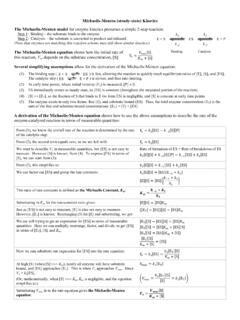
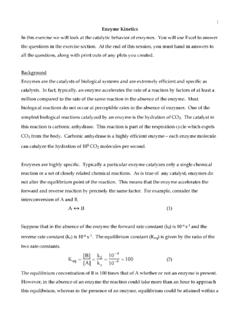
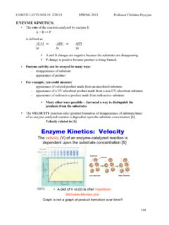
2 Enzyme Kinetics is the study of catalytic reactions, or reaction rate, which occurs in the presence of enzymes under varying conditions, specificities, and mechanisms such as the proximity effect, orientation effect, catalytic effect and energy effect; the studies are conducted under assorted circumstances, such as temperature, pH, and component concentrations in correlation to reaction rates. For example, Enzyme concentration directly relates to reaction rates whereby an increase in Enzyme concentration will also increase the rate of the reaction in a linear relationship. (as seen in the above graph on the left).
3 However, substrate concentration does not increase the reaction rate continuously if Enzyme concentration is constant, but rather reaches a maximum reaction rate or velocity known as Vmax when the Enzyme active sites are saturated with substrate and therefore can no longer bind to anymore substrate. The KM is the substrate concentration constant at half the Vmax value, in other words it is half of the maximum reaction rate which may aid in determination of Enzyme -substrate binding capabilities. The substrate concentration can be displayed as two differing graphs; the center graph located above, and as the Lineweaver Burk plot, also known as the double-reciprocal plot.
4 The Lineweaver Burk plot denotes KM and Vmax as intercept values in the form of reciprocals. The Vmax value is the point at which the plotted line intercepts the y-axis labeled 1/Vo interpreted as 1/Vmax. The KM value is determined at the point in which the plotted line intercepts with the x-axis labeled 1/[substrate], interpreted as -1/KM. The KM and Vmax values of obtained from the Lineweaver Burk plot may be used to determine reaction rate by use of Michaelis-Menton equation as follows: 2 Reaction Rate = Vo = (Vmax x [substrate]) / (KM + [substrate]) Other factors which affect reaction rates within Enzyme Kinetics include substrate specificity and temperature.
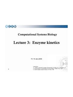
5 The effect of temperature on the reaction rate (as seen in the graph above on the right) of enzymes initially increases as temperature increases, until a maximum or optimal temperature is reached. At the optimal temperature, the highest reaction rate or catalytic activity occurs. Beyond the optimal temperature, the reaction rate rapidly decreases due to a temperature too high, above 50 to 60 oC, for the tertiary structure to withstand; the active site of the Enzyme is then disrupted due to unfolding of the structure and denaturation of the protein. Enzyme specificity toward substrate binding (as seen in the image below) is explained by two models, the lock-and-key model and the induced fit model.
6 The lock-and-key model was the original model used to explain the Enzyme -substrate complex fit whereby the enzymes and subtrates were thought to have rigid shapes which fit together as do a key into a lock, or two puzzle pieces; in this metaphor the Enzyme acts as the lock in which the substrate, as the key, must fit. However, the lock-and-key model has largely been replace by the induced fit model in which the Enzyme proceeds with a conformational change, or change in shape, at its active site around the substrate after the Enzyme -substrate complex formation. Methods: Part 1: Effe ct of Enzy me Concentration on Reaction Rate Assemble and label clean test tubes to prevent contamination or premature reaction from occurring.
7 Using a graduated cylinder, measure about 2 mL of the Enzyme , glucose oxidase at a concentration of units/mL and transfer to clean, labeled test tube. Measure about 4 mL of the substrate, M glucose and transfer to a separate clean and labeled test tube. In a separate beaker, prepare the reagent by combining 5 mL of horseradish peroxidase at a concentration of 40 units/mL, 5 mL of chromagen ( mM p-hydroxybenzoic acid + mM 4-aminoantipyrine), and 5 mL of M Tris-HCl pH buffer at pH Label 4 test tubes as samples 1 through 4, and distribute chemicals according to the following table: Test Tube Sample 1 2 3 4 Reagent Mixture (mL) Distilled Water (mL) units/mL Glucose Oxidase (mL) 3 Using a Vernier unit, place a sample of the reagent (in table above) into a vial within the colorimeter light source and tar to zero.
8 Return the sample from the vial to the test tube. Add 1 mL of M glucose to the test tube to initiate the reaction and immediately transfer to the vial within the light source to record absorbance values. Absorbance values are recorded for all 4 test tube samples at 468 nm (blue spectrum) per sample at 30 second intervals for seven minutes or until achieving an absorbance value of Part 2: Effe ct of Subs trate Concentration on Reaction Rate Assemble and label clean test tubes to prevent contamination or premature reaction from occurring. Using a graduated cylinder, measure about 4 mL of the Enzyme , glucose oxidase at a concentration of units/mL and transfer to clean, labeled test tube.
9 Measure about 2 mL each of the substrates, M glucose and M glucose, and transfer to separate clean and labeled test tubes. In a separate beaker, prepare the reagent by combining 10 mL of horseradish peroxidase at a concentration of 40 units/mL, 10 mL of chromagen ( mM p-hydroxybenzoic acid + mM 4-aminoantipyrine), and 10 mL of Tris-HCl pH buffer at pH Label 6 test tubes as samples 1 through 6, and distribute chemicals according to the following table: Test Tube Sample 1 2 3 4 5 6 Reagent Mixture (mL) Distilled Water (mL) units/mL Glucose Oxidase (mL) Using a Vernier unit, place a sample of the mixture in table above into a vial within the colorimeter light source and tar to zero.
10 Return the sample from the vial to the test tube. Add the quantity of substrate in the following table to the respective test tube sample to initiate the reaction: Test Tube Sample 1 2 3 4 5 6 M Glucose - - - M Glucose - - - Upon addition of the glucose substrate, the mixture is immediately transferred to the vial within the light source to record absorbance values. Absorbance values are recorded for all 6 test tube samples at 468 nm (blue spectrum) per sample at 30 second intervals for five minutes. Part 3A: Substrate Sp ecif ic ity Assemble and label 12 clean test tubes to prevent contamination or premature reaction from occurring.