Transcription of EYELID AND CANTHAL RECONSTRUCTION - vuyk
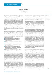
1 55 EYELID AND CANTHAL VuykINTRODUCTIONC onsidering the highly specialised multifunctional characteristics of the eyelids as wellas their aesthetic and emotional qualities, EYELID RECONSTRUCTION does have depth understanding of the anatomy together with the specific functional aspectsand aesthetic qualities of the EYELID is a prerequisite to adequately choose from an arrayof reconstructive surgical eyelids consist of various tissues layers (Fig. 1). At the level of the EYELID margin, thevarious layers from superficial to deep consist of skin, orbicularis, tarsus and the tarsal level, from superior to deep, additional tissue layers can be recognised,skin, orbicularis, orbital septum, periorbital fat, EYELID retractors and and orbicularis compose what is called the anterior lamella, while conjunctiva, lidretractors and tarsus are considered the posterior lamella24.
2 The tarsus consists not ofcartilage, but of dense, fibrous tissue and functions as stiffener. The height of the tarsalplate differs from upper and lower EYELID . In the upper EYELID the vertical dimensionranges from 9-12 mm, in the lower eyelidfrom 3-5 mm. The retractors attach to thefree border and slightly anterior portion ofthe tarsus. The retractors of the upper eye-lid consist of the levator aponeurosis andMuller s muscle. The capsulo-palpabralfascia in the lower EYELID is analogous tothe levator aponeurosis of the upper eyelidbut contains no muscle fibres. The capsulo-palpabral fascia transmits contractionsof the inferior rectus muscle while stabi-lising the tarsus of the lower , the retractors provide function,opposing the sphincteric action of theorbicularis muscle, as well as lid marginstability.
3 The orbital septum is a fascialmembrane separating the eyelids fromthe deeper orbital structures includingperiorbital 1. Anatomic layers of the medial CANTHAL complex constitutes the bony attachment of the eyelids, as well asthe lacrimal collecting and drainage system. Specifically the part of the medial canthaltendon which inserts posteriorly into the orbit helps maintain apposition of the eye-lids to the lateral CANTHAL area consists of 4structures, including the lateral canthaltendon, Lockwood s ligament, check liga-ments of the lateral rectus muscle, and thelateral horn of the levator , it encompasses the palpabrallobe of the lacrimal gland11.
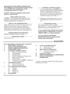
4 The lateralcanthal tendon attaches to Whitnall s tubercle, a bony promontory just withinthe bony rim. The lateral CANTHAL tendondraws the EYELID laterally, superiorly conceptualise EYELID reconstructionthe periorbital region is usually dividedin 4 zones. These zones are the following:Zone I upper EYELID ; Zone II lowereyelid; Zone III - medial canthus; ZoneIV lateral canthus7,11,17 (Fig. 2).FUNCTIONThe eyelids are a distinct facial feature that contribute to individual facial overall main purpose of the upper and lower EYELID and medial/lateral canthalcomplex, is support of adequate global function.
5 More specifically, the eyelids protectthe eyes from debris, excess light and exposure29. The upper EYELID is vastly more important to the function of corneal coverage than the lower eyelid33. Moreover, theupper EYELID provides lubrication by means of accessory lacrimal glands and ducts ofthe lacrimal gland. Untreated or inadequately treated defects of the upper EYELID willlead to severe dry eyes, resulting in corneal injury and visual loss. The lower eyelidwith its relatively stable margin, retains periglobal moisture, while blinking clears and redistributes the tearfilm. Both medial and CANTHAL tendon complex provide supportfor the EYELID and prevents malposition (entropion/ectropion) while maintaining adequate horizontal width of the palpable aperture.
6 The medial CANTHAL tendinouscomplex is part of the lacrimal drainage and tear pump system. If necessary, the lacri-mal drainage is probed and intubated for a long period to assure competence in me-dial CANTHAL defect RECONSTRUCTION . In its ideal position, both eyelids cover 1-2 mm ofthe limbus. On EYELID closure the upper EYELID must cover the cornea completely18, and CANTHAL reconstructionFig. 2. Periorbital zonesZone I upper eyelidZone IIlower eyelidZone III medial CANTHAL regionZone IV lateral CANTHAL region(Spinelli (1994) Periauricular RECONSTRUCTION : a systematic approach. Plast. Reconstr. Surg. 1 CONTROLE yelid skin is prone to skin cancer, and because it is so thin, most basal cell and squamouscell carcinomas invade the underlying orbicularis management of malignant tumours aim at disease eradication coupled withpreservation of adjacent normal tissue.)
7 In the periorbital region, functional and cosmeticsequelae of removal of excessive normal tissue may have far-reaching tumour ablative surgeon who is unfamiliar with the periorbital anatomy and reconstructive options, may compromise margins of resection because of the unwilling-ness to sacrifice vital structures in the micrographic surgery offers highest cure rates (< 98%) while preserving maximumamount of normal tissue in an area where excess tissue for RECONSTRUCTION is scarce1,4,6, invasion of basal cell carcinoma is rare but calls for extensive preoperativeevaluation and multidisciplinary team PRINCIPLES AND GUIDELINESMost resections and reconstructions are performed under local anaesthesia, sometimessupplemented with dissociative anaesthesia for enhanced patient comfort.
8 Localanaesthetic BMX drops are used in the conjunctival sac, while lidocaine 2% with ad-renaline 1 : is injected with a 30 G needle. For longer operations bupi-vacaine with epinephrine 1 : is added to the defects are generally categorized according to location (zone I-IV), depthand size. In anterior lamellar defects, part of the EYELID thickness remains. In fullthickness defects, both the anterior and posterior lamella must be reconstituted. Thelarger the percentage of the full thickness EYELID which is missing (0-25% / 25-50%/ > 50%) the more complex the ideal goal of RECONSTRUCTION is to provide global protection and normal to attain this goal may be the following:1.
9 Lining maintaining lubrication while avoiding corneal irritation2. Lid rigidity while allowig direct global apposition3. Lid stability and orientation with proper medial and lateral CANTHAL support4. Opening and closing ability facilitated by adequate muscle power and tone, allowedby subtle skin attain these goals, RECONSTRUCTION should ideally replace the delicate, thin, pliable, wellvascularised and innervated tissues of the eyelids with kind. In general, adjacent eyelidstructures may act as reservoirs for tissue harvesting and flap or graft transfer9,21,28,29, forces, such as gravity, edema and wound retraction may lead to com-plications, such as EYELID malposition (ectropion) and corneal and CANTHAL reconstructionAnterior lamellar defectsconsisting of partial EYELID thickness are generally accomplishedwith local flaps or full thickness grafts28.
10 Most local flaps consist of skin and muscularisto enhance vascularity, replace bulk and possibly provide active muscle tonus to supportthe EYELID function. The tissue reservoirs to be considered for flap harvesting are uppereyelid, temple/cheek and glabella region for lower EYELID defects and temple/suprabrowand glabellar region for upper eyelid5. Alternatively, grafts from the ipsilateral or contralateral EYELID or composite skin-perichondrium grafts of the periauricular regionmay be applied in both upper and lower EYELID reconstruction28, upper EYELID presents an obvious reservoir of redundant skin and muscle to beharvested for skin-muscle thickness composite skin-orbicularis-fascia grafts from the upper EYELID may beversatile in RECONSTRUCTION anterior lower and upper EYELID defects28.