Transcription of Fetal Pig Dissection Unit - Grosse Pointe Public …
1 1 Name _____ Date _____ Hour ____ Fetal Pig Dissection Unit Objectives: Identify important external structures of the Fetal pig. Identify major structures associated with a Fetal pig's integumentary, skeletal, digestive, respiratory, circulatory, urogenital, & nervous systems. Compare the functions of certain organs in a Fetal mammal with those of an adult mammal. Materials: preserved Fetal pig, dissecting pan, dissecting tools, dissecting T-pins, plastic bag, metric ruler, paper towels, latex gloves, string General Dissection Information: The term Dissection means more than merely cutting your specimen apart.
2 It is a method of seeking, exposing, identifying and studying the internal anatomy. It also helps to bring into view many structures not readily seen under normal circumstances. Remember the Dissection tools are sharp and are to be handled with caution and care. Always clean these tools after using them. Use your scissors to do the cuts. Rely mostly on the probes for looking at and pushing aside internal structures. When studying a system or organ, always look at the diagram, making sure of the name and location of the body part. Greater caution must be taken in dissecting a Fetal animal than an adult animal.
3 The organs and tissues are not yet fully developed. They are, therefore, smaller, differently shaped, and more delicate than those of the adult. It will mean using greater care in cutting the soft, thin skin. A careless movement of finger, scissor, or probe may tear, cut, or destroy important structures. At the end of each class period, wrap the Fetal pig in wet paper toweling, wet it and put it in the plastic bag. Do not allow for much air in the bag, for this may cause the Fetal pig to decay prematurely. Remember to line your Dissection pan with paper toweling before placing the specimen on it.
4 This will help your clean up! When all supplies are cleaned and properly put away, wash your hands with soap and water. 2 Background Information: The Fetal pig (Sus scrofa) belongs to the class Mammalia , the same class to which man belongs. The gestation period of the pig is about 115 days and the Fetal pigs are approximately 30 cm in length at the end of this period. Mammals are vertebrates having hair on their body and mammary glands to nourish their young. The majority are placental mammals in which the developing young, or fetus, grows inside the female s uterus while attached to a membrane called the placenta.
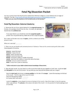
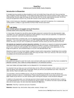
5 The placenta is a source of food and oxygen for the fetus, and it also serves to get rid of Fetal wastes. The Dissection of the Fetal pig in the laboratory is important because pigs and humans have the same level of metabolism and have similar organs and systems. ---------------------- External Anatomy 1. Obtain a Fetal pig and rinse off the excess preservative by holding it under running water. Lay the pig on its side in the dissecting pan and locate the dorsal (back surface), ventral (belly surface), anterior (toward the head), and posterior (toward the tail end) surfaces.
6 2. A Fetal pig has not been born yet, but its approximate age since conception can be estimated by measuring its length. Measure your pig's length from the tip of its snout to the base (start) of its tail, using string. Do not measure the tail itself. **Use the length/age chart below to determine the age of your Fetal pig and record this on your hand-in for day 1. Length of Fetus Gestation Days 2 cm 35 days 4 cm 56 days 8 cm 68 days 10 cm 74 days 12 cm 80 days 14 cm 86 days 16 cm 92 days 22 cm 100 days 30 cm 112-115 days 3 External Structures a.
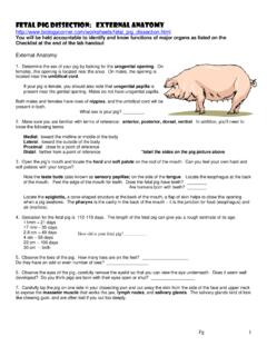
7 Snout The snout of the pig has a blunt tip composed of cartilage and strengthened by bone. This permits the pig to use the snout to push, lift weights, and dig. b. Nares These are the nostrils of the snout. They open into the nasal cavity. Here the inhaled air is warmed, filtered, and moistened. c. Pinna This is the flap-like external ear. They are composed of cartilage, just as the human ear is composed. Its function is to collect sound waves. d. Eyes Note the upper and lower eyelids. In addition, there is a third eyelid, the nictitating membrane, which covers the anterior portion of the eye and helps to clean it.
8 **You need to make a small incision and open the other two lids to see this. e. Appendages Note the positions of the knee, the elbow, the ankle, and the wrist. f. Digit (on the foot) The pig walks on the digits, or toes. **Count and record the number of digits of the Fetal pig. g. Umbilical cord This extends from the abdomen of the fetus to the placenta of the mother. It functions in the bringing of food and oxygen to the fetus and the removal of wastes. Substances diffuse from one blood system to the other. ** With scissors, cut across the cord about 1 cm from the body.
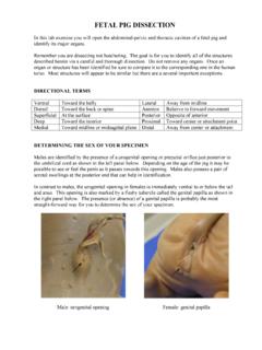
9 Examine the 3 openings in the umbilical cord. The largest is the umbilical vein, which carries blood from the placenta to the fetus. The two smaller openings are the umbilical arteries which carry blood from the fetus to the placenta. h. Anus - Lift the pig's tail to find the anus. Study the ventral surface of the pig and note the tiny bumps called mammary papillary. These are present in both sexes. In the female these structures connect to the mammary glands. i. Urogenital Opening - Determine the sex of your pig by locating the urogenital opening through which liquid wastes and reproductive cells pass.
10 In the male, the opening is on the ventral surface of the pig, just posterior to the umbilical cord. In the female, the opening is ventral (just below) to the anus. **Record the sex of your pig. male female 4 The Skeleton The skeleton of the Fetal pig has not yet fully hardened to bone. Much of the skeleton is composed of cartilage. Skeletal Structures: a. Feet - Observe the position of the feet in the diagram of the adult pig. While man walks on the sole of the foot, the pig walks on his toes. The feet are narrow and foot bones are separate. The 1st digit of the pig is absent.