Transcription of FOCUS Sports injuries Sports ankle injuries - Peter …
1 FOCUS Sports injuries Sports ankle injuries AUTHOR: PLEASE. HEAD AND. SHOULDER. Assessment and management IMAGE. Drew Slimmon Peter Brukner Background Case study Lucia is a female, 16 years of age, who plays Sports ankle injuries present commonly in the general netball with the state under 17s netball practice setting. The majority of these injuries are inversion team. She presents with an ankle injury and plantar flexion injuries that result in damage to the sustained at training the previous night. She lateral ligament complex. is on crutches and is nonweight bearing. Objective Examination raises the possibility of a The aim of this article is to review the assessment and fracture, but X-ray is negative. You diagnose management of Sports ankle injuries in the general practice a severe lateral ligament sprain and manage setting. Lucia with ice, a compression bandage and Discussion a backslab initially. She then progresses Assessment of an ankle injury begins with a detailed history through a 6 week rehabilitation program and to determine the severity, mechanism and velocity of the you recommend she wear an ankle brace for injury, what happened immediately after and whether at least 6 months.
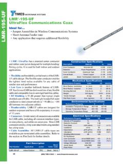
2 There is a past history of inadequately rehabilitated ankle injury. Examination involves assessment of weight bearing, inspection, palpation, movement, and application of special examination tests. Plain X-rays may be helpful to exclude a fracture. If the diagnosis is uncertain, consider second The majority of ankle injuries are inversion and plantar line investigations including bone scan, computerised flexion injuries that result in damage to the lateral tomography or magnetic resonance imaging, and referral to a ligament complex (Figure 1). The main ligaments of Sports physician. Manage all lateral ligament complex ankle concern are the anterior talofibular ligament (ATFL), sprains with ice, compression, elevation where possible and the calcaneofibular ligament (CFL), and the anterior analgesia. Severe ligament sprains or rupture benefit from tibiofibular ligament (also called the anterior inferior a brief period of immobilisation. After initial management, tibiofibular ligament or AITFL).
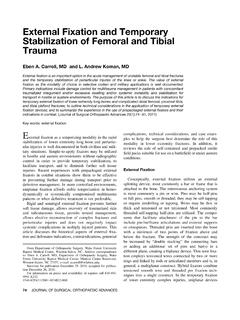
3 The AITFL is the anterior the athlete should complete a 6 week guided rehabilitation component of the syndesmosis complex (Figure 2). A. program. Athletes with moderate to severe lateral ankle thorough history including past history and examination ligament sprains should wear a semirigid or rigid ankle orthosis for at least 6 months following injury. will determine the need for investigations and guide appropriate management. Keywords: ankle ; athletic injuries ; soft tissue injuries History Injury details Severe ligament sprains or ruptures occur with high velocity injuries , such as landing on a player's foot while jumping or running. Simply rolling the ankle on an uneven surface while walking is unlikely to cause a ligament rupture or fracture in the setting of normal bone density and ligament integrity, ie. there is no history of prior ligament damage. Dorsiflexion and eversion injuries can cause damage to the syndesmosis (Figure 2). A syndesmosis injury takes significantly longer to recover than a lateral ligament injury and, if unstable, warrants immediate surgical referral.
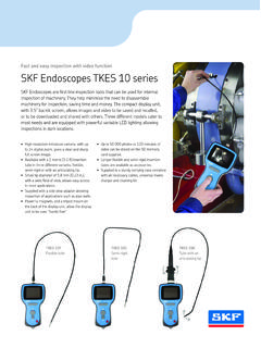
4 18 Australian Family Physician Vol. 39, No. 1/2, January/February 2010. Post injury details Lateral talar tilt test After a ruptured ligament or fracture, athletes are unlikely to be able This is performed by applying an inversion force to the ankle joint at to continue training or playing sport, and may be unable to weight the heel, then assessing the degree of talar tilt. A talar tilt of greater bear (particularly with a fracture). Swelling within minutes or hours than 15 degrees reflects rupture of the ATFL and CFL (Figure 4).3,4. is the result of the bleeding that occurs with a ligament rupture or fracture/dislocation; swelling from synovitis takes longer to develop. Reports of a cracking noise or the feeling that the ankle bent Posterior tibiofibular double cannot differentiate between a fracture, ligament rupture or ligament Anterior tibiofibular ligament ligamentous Posterior Anterior talofibular ligament Past history talofibular ligament Bifurcate ligament The past history provides the practitioner with an understanding of the state of the ankle before the current injury.
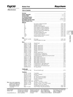
5 Inadequate rehabilitation and a subsequently chronically unstable ankle would be expected to take significantly longer to completely rehabilitate. It is important Calcaneofibular ligament Long plantar ligament to clarify the extent of any previous injury and any investigations or treatment. If a rehabilitation program was instituted, the clinician Figure 1. Lateral ankle ligaments should seek details of the type and duration of the program. Appropriate Reproduced from Brukner P, Kahn K. Clinical Sports medicine, rehabilitation programs FOCUS on range of motion, proprioception, 3rd edn. Sydney: McGraw-Hill, 2007. strength and Sports specific exercises, and are usually a minimum of 6 weeks duration. Asking about previous or current use of a semirigid brace or tape may offer insight into the adequacy of past rehabilitation efforts as this is an important component of rehabilitation. Interosseous Examination membrane Examination of the injured ankle includes: Weight bearing if the patient cannot weight bear on the affected Syndesmosis leg for more than four steps unaided, ankle X-rays should be tear ordered (Figure 3)2.
6 Inspection discoloration hematoma and significant swelling are suggestive of ligament rupture or fracture Anterior Palpation pain on palpation of the posterior aspect of the tibiofibular ligament medial and lateral malleolus (both sites are void of ligamentous attachments), the base of the fifth metatarsal and the proximal fibula are suggestive of fracture (Figure 3).2 Lack of tenderness on palpation of the ATFL excludes ATFL rupture Range of movement this may not assist diagnosis acutely and is likely to be limited by acute swelling and pain. Special examination tests These may not assist diagnosis acutely as a result of swelling and pain. In this situation the examination should be repeated 3 5 days later when pain and swelling have subsided. Anterior drawer test This is performed with the knee at 90 degress of flexion and the muscles relaxed. Increased laxity compared to the contralateral ankle reflects injury to the ATFL and possibly the CFL. The combination of Figure 2. Syndesmosis sprain discoloration hematoma, ATFL tenderness and positive anterior drawer Reproduced from Brukner P, Kahn K.
7 Clinical Sports medicine, 3rd edn. provides sensitivity of 98% and a specificity of 84% of an acute lateral Sydney: McGraw-Hill, 2007. ligament Australian Family Physician Vol. 39, No. 1/2, January/February 2010 19. FOCUS Sports ankle injuries assessment and management Figure 3. Ottawa ankle rules [AUTHOR: DO YOU HAVE PERMISSION TO REPRODUCE THIS?]. Tests for a syndesmosis injury External rotation stress test, squeeze test and interosseous membrane tenderness length should be performed if the mechanism suggests a syndesmosis injury, or if there is tenderness on palpation of the AITFL Figure 4. Lateral talar tilt test (Table 1). Importantly, following an ankle inversion plantarflexion injury, 60% of patients will have pain over the medial malleolus in the absence of a syndesmosis injury or medial malleolus Consider the commonly missed fractures around the ankle (Table 2). This is also an opportune time to consider referral to a Sports physician. Investigations While a plain X-ray excludes a fracture in the vast majority of Plain X-rays are often unnecessary in the case of uncomplicated lateral cases, a bone scan or computerised tomography (CT) scan may be ligament complex injuries .
8 The Ottawa ankle Guidelines (Figure 3) helpful if an occult fracture is suspected. These investigations are provide a well validated guide to assessing whether an X-ray is required relatively inexpensive compared to magnetic resonance imaging to exclude a Clinical assessment is usually adequate initially (MRI) and are highly sensitive for bony pathology. Focally increased to assess the severity of ligament damage. There is no evidence based radioisotope uptake on the delayed phase of a bone scan confirms role for stress radiographs or ultrasound in acute ankle injuries . bone damage; CT provides further anatomical detail and assists If the patient fails to improve significantly after a well guided determining whether surgery is required. rehabilitation period of at least 6 weeks then they should be reassessed Magnetic resonance imaging can be useful as a second line and the differential diagnosis reviewed. At this stage, plain radiographs investigation, usually in the specialist setting.
9 It can clarify diagnoses may be reconsidered to exclude an occult osteochondral lesion. of ligament sprain or rupture, reveal ongoing synovitis, and assess osteochondral damage that may require surgical referral. Table 1. Clinical testing for syndesmosis injury Diagnosis and management External rotation The patient's ankle is passively For management purposes, ankle injuries can be considered under the stress test dorsiflexed in maximal external rotation following headings: (either seated or lying prone with knee flexed to 90 degrees). Pain at the Mild to moderate lateral ligament complex sprain treatable with syndesmosis is regarded as a positive test early mobilisation and guided proprioceptive and strengthening Squeeze test With both hands clasp the medial rehabilitation program and lateral aspects of the midcalf and Severe lateral ligament complex sprain immobilisation for a squeeze. Pain distally at the site of the period of days in a cast (a simple backslab is sufficient) or cam syndesmosis is regarded as a positive walker (walking boot) followed by a guided proprioceptive and test strengthening rehabilitation program Interosseous Patient sits or lies supine with their Fracture/dislocation or unstable syndesmotic injury requiring membrane affected leg extended on examination tenderness length table.
10 Palpate between the fibula and orthopedic surgeon referral. tibia from the ankle joint proximally. Acute phase management of lateral Determine the length of tenderness from the distal tip of the fibula and ligament complex strains document Once a fracture/dislocation or unstable syndesmotic injury is excluded, 20 Australian Family Physician Vol. 39, No. 1/2, January/February 2010. Sports ankle injuries assessment and management FOCUS . Table 2. Commonly missed fractures compression alone or an Severe was defined as being unable to weight bear for at least 3 days without radiological Proximal fibula evidence of fracture. It is now generally accepted that following a Base of fifth metatarsal severe ligament sprain or rupture, initial protected immobilisation Anterior process of calcaneus is likely to be beneficial. A backslab or cam walker usually provides Lateral talar process adequate protection. A more diagnostic examination is usually Posterior process of talus (also os trigonum fracture) possible when pain and swelling have decreased and following Talar dome a period of immobilisation the patient can progress to a graded Tibial plafond rehabilitation program.