Transcription of For Medial High Tibial Osteotomies TomoFix™ …
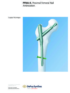
1 For Medial high Tibial Osteotomies tomofix Medial high Tibial Plate (MHT). surgical Technique Image intensi er control This description alone does not provide sufficient background for direct use of DePuy Synthes products. Instruction by a surgeon experienced in handling these products is highly recommended. Processing, Reprocessing, Care and Maintenance For general guidelines, function control and dismantling of m ulti-part instruments, as well as processing guidelines for i mplants, please contact your local sales representative or refer to: For general information about reprocessing, care and maintenance of Synthes reusable devices, instrument trays and cases, as well as processing of Synthes non-sterile implants, please consult the Important Information leaflet (SE_023827) or refer to: Table of Contents Introduction Indications and Contraindications 4. General Remarks 5.
2 surgical Technique Open Wedge surgical Technique 6. Preparation and Approach 6. Osteotomy 13. Positioning and Fixation of the Plate 33. Postoperative Treatment and Implant Removal 51. Closed Wedge surgical Technique 52. Preparation 52. Osteotomy 56. Product Information Plates 58. Screws 60. Kirschner wires 61. Instruments 62. Optional Instruments 65. Cases 66. Optional Case 67. Also Available from DepuySynthes: chronOS 68. MRI Information 69. Bibliography 70. tomofix Medial high Tibial Plate (MHT) surgical Technique DePuy Synthes 1. tomofix Medial high Tibial Plate (MHT). For Medial high Tibial Osteotomies Features Four locking screws in the plate head Long shaft portion evenly maintain stability transmits the occurring forces into the Tibial shaft Compression of the lateral hinge A lag screw pulls the distal osteot- and forces the plate into suspen- which imposes pressure upon the omy segment towards the plate sion, creating an elastic preload lateral hinge.
3 2 DePuy Synthes tomofix Medial high Tibial Plate (MHT) surgical Technique Tapered, rounded tip facilitates plate insertion. Pretensioning of the plate allows compression of the lateral hinge tomofix Knee Osteotomy System tomofix Tibial Head tomofix Tibial Head tomofix Femoral Plate tomofix Femoral Plate Plate Medial , proximal Plate lateral, proximal Medial , distal lateral, distal For open and closed For open and closed For closed-wedge osteo For open and closed wedge high Tibial osteo wedge osteo tomies tomies wedge Osteotomies tomies Fixed-angle construct for Fixed-angle construct for Fixed-angle construct vor Allows application of the stable fixation stable fixation stable fixation preload technique Available in right and left Available in right and left Available in right and left Provides support for versions versions versions stable bridging*. Available in standard, small and anatomical stature versions * Mechanical testing data on file.
4 tomofix Medial high Tibial Plate (MHT) surgical Technique DePuy Synthes 3. Indications and Contraindications Indications Open-wedge and closed-wedge Osteotomies of the Medial proximal tibia for the treatment of: Unicompartmental Medial or lateral gonarthrosis with malalignment of the proximal tibia Idiopathic or posttraumatic varus or valgus deformity of the proximal tibia Contraindications Inflammatory arthritis 4 DePuy Synthes tomofix Medial high Tibial Plate (MHT) surgical Technique General Remarks high Tibia Osteotomy is becoming an increasingly popu- lar method to treat unicompartmental OA of the knee. This joint preserving procedure plays a critical role within the continuum of care, as when performed precisely, it can delay or eliminate the need for joint replacement. The tomofix knee osteotomy system is based on the Locking Compression Plate system (LCP) and enables angular-stable connections between the screw and plate.
5 This angular stability allows the stable fixation of an osteotomy intended for early and safe mobilization in accordance with the AO principles. Note: Plan the type and position of the osteotomy. The tomofix Medial high Tibial Plate is suitable for both open and closed-wedge Osteotomies . This surgical technique will explain the procedure of an open and closed wedge osteotomy. For information on transverse and sagittal plane Osteotomies please consult Osteotomies around the knee by Lobenhoffer P, RJ. van Heerwaarden, AE Staubli, RP Jakob (see Bibliogra- phy on page 70). tomofix Medial high Tibial Plate (MHT) surgical Technique DePuy Synthes 5. Open Wedge surgical Technique Preparation and Approach 1. Preoperative Planning 1. A precise preoperative plan is crucial to the success of this procedure. The recommended method for planning is that of Miniaci. It must be done on the basis of the weight-bearing x-ray of the full leg in AP view, either on paper or at a digital workstation.
6 Determine the mechanical axis of the leg: Draw a straight line from the center of the femoral head to the center of the ankle joint. h h o Draw the new weight-bearing line from the center of the femoral head, passing the joint line through the desired position. Determine a hinge point (h). Generally the hinge point should be chosen on the lateral cortex and at the up- per 1/3 proximal fibular head. Note: The optimal position of the hinge point may ba a b a vary according to patient specific anatomy. Rotate the leg 30 internally to identify the optimal 2. hinge point. The lateral hinge point should be within the proximal 1/3 of the fibular head. (Han et al., see Bibliography ). Connect the hinge point (h) with the center of the ankle joint (a). Rotate the connecting line h-a like a cir- cle until it crosses the new weight bearing line. Con- nect the crossing point (b) with the hinge point h.
7 The angle between the connecting line h-a and h-b is the angle of opening (a). Transfer the opening angle (a) to the level of the planned osteotomy. The height at the Medial cortex (o) is the height of opening. (1). Note: If the height is measured intraoperative it should be calculated as height of opening plus thick- ness of the saw blade ( mm). 6 DePuy Synthes tomofix Medial high Tibial Plate (MHT) surgical Technique Determine the entry point of the transverse osteotomy. It lies just above the pes anserinus. Make sure there is still enough space for the proximal part of the tomofix plate (holes A-D), so that the screw in hole D can be inserted without protruding into the opening gap. Depending on the determined opening angle and the length of the osteotomy cut (mediolateral diameter of the osteotomy). the corresponding opening height can be derived from Hernigou's trigonometric chart.
8 Trigonometric chart Correction Angle 4 5 6 7 8 9 10 11 12 13 14 15 16 17 18 19 . 50 mm 3 4 5 6 7 8 9 10 10 11 12 13 14 15 16 16. Mediolateral diameter of the osteotomy (mm). 55 mm 4 5 6 7 8 9 10 10 11 12 13 14 15 16 17 18. 60 mm 4 5 6 7 8 9 10 11 12 14 15 16 17 18 19 20. 65 mm 5 6 7 8 9 10 11 12 14 15 16 17 18 19 20 21. 70 mm 5 6 7 8 10 11 12 13 15 16 17 18 20 21 22 23. 75 mm 5 6 8 9 10 12 13 14 16 17 18 20 21 22 24 25. 80 mm 6 7 8 10 11 13 14 15 17 18 19 21 22 24 25 26. Note: These instructions alone do not replace in- depth training in planning for Osteotomies and only serve as a general guideline. tomofix Medial high Tibial Plate (MHT) surgical Technique DePuy Synthes 7. Open Wedge surgical Technique Preparation and Approach 2. Prepare the implant Instruments and implants Guiding Block for tomofix Tibial Head A. Plate, small, Medial , proximal B. and C. tomofix Tibial Head Plate, small, Medial , proximal, shaft 4 holes, head 4 holes, length 112 mm, A.
9 Pure Titanium, sterile B. or C. tomofix Guiding Block for tomofix Tibial Head Plate, Medial , proximal 4 3 2 1 D. and C. tomofix Tibial Head Plate, Medial , B. proximal, 4 holes, Pure Titanium, sterile A. or tomofix Guiding Block for tomofix Tibial Head Plate, anatomical, proximal, A. Medial , left B. and C. tomofix Tibial Head Plate, anatomical, Medial , proximal, left, head 4 holes, length 112 mm, Pure Titanium, sterile or tomofix Guiding Block for tomofix Tibial Head Plate, anatomical, proximal, Medial , right and tomofix Tibial Head Plate, anatomical, Medial , proximal, right, head 4 holes, length 112 mm, Pure Titanium, sterile LCP Drill Sleeve , for Drill Bits B mm LCP Spacer B mm, length 2 mm, Titanium Alloy (TAN). 8 DePuy Synthes tomofix Medial high Tibial Plate (MHT) surgical Technique Choose the corresponding guiding block for either the standard, small or anatomical tomofix plate.
10 Due to the shape of the anatomical plate there is a left and right version. The guiding block is marked with L or R. accordingly. Place the guiding block on the plate. The guiding block serves as an aid for attaching the LCP drill guides at the correct angle and should be removed after the drill guides have been attached. Screw in and tighten a LCP drill guide into holes A, B. and C. Insert a LCP spacer B mm into hole D and hole 4. Notes: Using spacers allows for the pes anserinus to move freely underneath the plate as well as for bending of the plate. This creates a tension that will act on the lateral hinge, thus generating compression. The anatomical tomofix plates ( , ) are provided precontoured and should not be bent prior to implantation. tomofix Medial high Tibial Plate (MHT) surgical Technique DePuy Synthes 9. surgical Technique Open Wedge Preparation and Approach 3.
