Transcription of FUNCTIONS OF THE NERVOUS SYSTEM
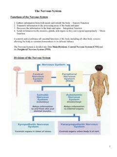
1 FUNCTIONS OF THE NERVOUS SYSTEM . 1. Sensory input. Sensory receptors detects external and internal stimuli. 2. Integration. The brain and spinal cord process sensory input and produce responses. 3. Homeostasis. The NERVOUS SYSTEM coordinates the activities of other systems in order to maintain homeostasis. 4. Mental activities. "I say to myself, Self? What do you think?". 5. Controls muscles and glands. DIVISIONS OF THE NERVOUS SYSTEM . FIGURE 1. The NERVOUS SYSTEM can be divided into subdivisions based on structure and on function. Each of the these subdivisions are referred to as separate NERVOUS systems. However, keep in mind that the subdivisions are all part of a single NERVOUS SYSTEM .
2 2. Central NERVOUS SYSTEM (CNS). A. The CNS consists of the brain and spinal cord. B. The CNS is enclosed by bone (the skull and vertebrae). 3. Peripheral NERVOUS SYSTEM (PNS). A. The PNS consists of sensory receptors, nerves, ganglia, and plexuses. B. Sensory receptors are the endings of nerve cells or separate, specialized cells. They detect stimuli, such as temperature, touch, pain, etc. C. Nerves are bundles of axons that connect the CNS to sensory receptors, muscles, and glands. Nerves are divided into two groups. 1) Cranial nerves. Twelve pairs arise from the brain and exit through foramina in the skull. 2) Spinal nerves.
3 Thirty one pairs arise from the spinal cord and exit through the intervertebral or sacral foramina. D. A ganglion is a collection of neuron cell bodies in the PNS. E. A plexus is an extensive network of axons, and in some cases neuron cell bodies, in the PNS. 11-1. 4. The PNS can be divided into two parts. FIGURE A. The sensory, or afferent, division carries action potentials TO the CNS. 1) Sensory input can be at the conscious level, such as touch, taste, smell, sight, etc. 2) Sensory input can be at the unconscious level, such as blood pressure, blood oxygen levels, etc. B. The motor, or efferent, division carries action potentials FROM the CNS to effector organs and can be divided into two parts.
4 1) The somatic NERVOUS SYSTEM innervates skeletal muscle. It is usually under voluntary conscious control, although reflexes are involuntary. 2) The autonomic NERVOUS SYSTEM (ANS) innervates smooth muscle, cardiac muscle, and glands. It is usually under involuntary control, although biofeedback and meditation techniques can produce voluntary control of normally involuntary FUNCTIONS . The ANS can be divided into three parts. a. The sympathetic division prepares the body for physical activity. b. The parasympathetic division maintains normal resting FUNCTIONS , such as digestion of food. c. The enteric NERVOUS SYSTEM consists of plexuses in the wall of the digestive tract.
5 Although it has sympathetic and parasympathetic neurons, it has its own neurons and can function independently of the CNS. 5. The central NERVOUS SYSTEM receives input from the PNS, integrates the input and causes a response. The CNS is the integrative and control center of the NERVOUS SYSTEM . The peripheral NERVOUS SYSTEM receives stimuli from the environment and conducts action potentials to and from the CNS. FIGURE CELLS OF THE NERVOUS SYSTEM . 1. Neurons receive stimuli and conduct action potentials. 2. Neuroglia are support cells that perform a variety of FUNCTIONS . Neuroglia cells account for over half the brain's weight.
6 There can be 10 to 50 times more neuroglia cells than neurons in various locations in the brain. Neurons FIGURE 1. The neuron cell body contains the nucleus, and the cell body is the site of manufacture of proteins (enzymes) necessary for neuron function. 11-2. 2. Dendrites are short, cytoplasmic extensions from the neuron cell body. They are specialized to receive stimuli, which can result in the production of an action potential in the neuron. 3. Axons, or nerve fibers, are long cytoplasmic extensions from the neuron cell body. A. The axon arises from an enlarged area of the neuron cell body called the axon hillock.
7 The beginning of the axon is called the initial segment. The trigger zone consists of the axon hillock and initial segment. It is where action potentials are generated. B. Axons are specialized to conduct action potentials to their ends, the presynaptic terminals. Neurotransmitters are chemicals released from the presynaptic terminals. Types of Neurons FIGURE 1. There are three types of neurons based on their structure. A. Multipolar neurons have several dendrites and one axon. B. Bipolar neurons have one dendrite and one axon. C. Unipolar neurons have one axon. 1) During development unipolar neurons were originally bipolar neurons.
8 2) The dendrite and axon migrated around the cell body and fused together. 3) Usually the fused processes divide into two processes that are morphologically identical to axons, so both processes are called axons. 2. Types of neurons according to function. A. Sensory neurons transmit action potentials TO the CNS. Most are unipolar, but a few sensory neurons associated with the senses of sight, taste, hearing, and smell are bipolar. B. Motor neurons transmit action potentials AWAY FROM the CNS. They are multipolar. C. Interneurons transmit impulses from one neuron to another neuron. They are multipolar. Neuroglia of the CNS.
9 Astrocytes FIGURE 1. Astrocytes have extensions that cover the surfaces of blood vessels and neurons. 2. Astrocytes function as a nonrigid supporting matrix for blood vessels and neurons in the brain. 11-3. 3. Astrocytes help to regulate the composition of the extracellular fluid around neurons. A. Astrocytes remove and/or process materials that pass through blood vessels into the extracellular fluid. B. Astrocytes influence the formation of tight junctions between the epithelial cells of blood vessels. The epithelial cells form the blood-brain barrier, which controls what substances can enter the central NERVOUS SYSTEM .
10 Ependymal Cells FIGURE 1. Ependymal cells line the cavities of the brain and spinal cord. 2. In specialized areas called the choroid plexuses they produce cerebrospinal fluid. Microglia FIGURE 1. Microglia are phagocytic cells in the CNS. 2. They engulf microbes or damaged tissue. Oligodendrocytes FIGURE 1. Oligodendrocytes surround axons in the central NERVOUS SYSTEM . 2. If the plasma membrane of an oligodendrocyte wraps many times around an axon, it forms a myelin sheath. Analogy: the axon is the cardboard center of a role of toilet paper, and the plasma membrane is the toilet paper. 3. The plasma membrane of an oligodendrocyte forms a myelin sheath around more than one axon.