Transcription of Genetics of human iris colour and patterns - …
1 Pigment Cell Melanoma Res. 22; 544 562 REVIEW ARTICLE. Genetics of human iris colour and patterns Richard A. Sturm1 and Mats Larsson2,3. 1 Melanogenix Group, Institute for molecular Bioscience, The University of Queensland, Brisbane, Qld, Australia KEYWORDS Eye colour melanocyte OCA2 . 2 Queensland Institute of Medical Research, Brisbane, Qld, Australia and 3 Center for Developmental Research, HERC2 quantitative trait transcription factor . School of Law, Psychology and Social Work, rebro University, rebro, Sweden gene regulation MITF PAX6. CORRESPONDENCE R. A. Sturm, e-mail: PUBLICATION DATA Received 27 May 2009, revised and accepted for publication 6 July 2009. doi: Summary The presence of melanin pigment within the iris is responsible for the visual impression of human eye colour - ation with complex patterns also evident in this tissue, including Fuchs' crypts, nevi, Wolfflin nodules and contraction furrows.
2 The genetic basis underlying the determination and inheritance of these traits has been the subject of debate and research from the very beginning of quantitative trait studies in humans. Although segregation of blue-brown eye colour has been described using a simple Mendelian dominant-recessive gene model this is too simplistic, and a new molecular genetic perspective is needed to fully understand the biolog- ical complexities of this process as a polygenic trait. Nevertheless, it has been estimated that 74% of the variance in human eye colour can be explained by one interval on chromosome 15 that contains the OCA2. gene. Fine mapping of this region has identified a single base change rs12913832 T C within intron 86 of the upstream HERC2 locus that explains almost all of this association with blue-brown eye colour .
3 A model is presented whereby this SNP, serving as a target site for the SWI SNF family member HLTF, acts as part of a highly evolutionary conserved regulatory element required for OCA2 gene activation through chromatin remodelling. Major candidate genes possibly effecting iris patterns are also discussed, including MITF and PAX6. relatively melanin-free layers, collagen fibrils of the iris Introduction scatter the short blue wavelengths to the surface, thus The study of human eye colour as a physical trait is a blue iris is a consequence of structure not of major based on the developmental biology, morphology, differences in chemical composition. Different shades chemistry and genetic determinants of the structure of blue, and in irises with a limited amount of melanin, known as the iris , which is part of the uveal tract of different shades of grey, green and hazel, are deter- the eye (Forrester et al.)
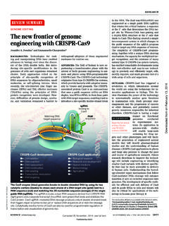
4 , 2008). The iris is a small con- mined by the thickness and density of the iris itself nective tissue and muscular structure of around and the extent of accumulation of white collagen 12 mm in diameter with a central opening called the fibres, as well as patches of tissue loss in the anterior pupil (Figure 1). It controls the amount of light entering border layer and stroma. the eye which is focused by the lens onto the retina However, a careful examination of people's irises so as to provide the sense of vision. It contracts in makes it clear that there are characteristics other than bright light making the pupil smaller and dilates in dark eye colour that present in the human iris . The iris has conditions making the pupil larger, which together with been analysed to show that it can display a degree of the source of the incident light can influence the per- complexity encompassing over 240 degrees of freedom ception of an individual's eye colour and iris pattern (Daugman, 2003), and is probably the most complex tis- (Figure 2).
5 In the brown iris there is an abundance of sue structure that is readily available on the outside of melanocytes and melanin in the anterior border layer the human body. iris tissue forms complex patterns that and stroma whereas in the blue iris these layers contain many distinct features (Figure 2B), which are contain very little melanin. As light traverses these currently used as biomarkers for a wide variety of 544 2009 John Wiley & Sons A/S. iris colour and patterns A B. Figure 1. human iris structure. (A) Frontal sector of the iris is shown with the collarette separating the papillary and ciliary zones. (B) A transverse view illustrating the five layers found within the iris . different purposes. For example, the tissue patterns extremity of the optic cup (Eagle, 1988).
6 In contrast to that are present in the human iris can serve as a reliable the IPE, the stromal melanocytes are of the same basis for automatic personal identification. An increasing embryological origin as the dermal melanocytes, in the number of banks use the iris as a mean of identification neural crest, and migrate through the uveal tract during rather than pin codes. At least 1 million frequent travel- development. The IPE is always pigmented in the exam- lers to the UK will be able to enter the country without ination of the iris in all eye colours, except in individuals presenting a passport or explicitly asserting their iden- with albinism, and contributes little to the impression of tity. Instead, their iris patterns are captured by a video eye colour .
7 Where the above lying stroma is thin this camera for comparison against a database of authorized layer can have some influence on patterning. For exam- persons (Daugman, 2006). Furthermore, tissue markers ple, the dark colouration of the IPE can absorb the pene- in the iris are associated with eye diseases such as ocu- trating light in eyes with a very thin stroma, which will lar melanoma, glaucoma (Chang et al., 1999), pigment give the white collagen fibres in the deeper cell layers dispersion and pseudoexfoliation syndrome (Asano of the stroma a grey tinge (Figure 2B, panel 1). The IPE. et al., 1995), as well as neurological disease such as may serve a protective role for the retina by absorbing Downs Syndrome (Donaldson, 1961), Neurofibromatosis excess light that can only enter through the pupil and is type 1 (Lee and Stephenson, 2007) and Gillespie syn- not able to be reflected back out.
8 Non-pigmented drome (Marien et al., 2008; Ticho et al., 2006), which smooth muscle differentiated tissue at the stromal-IPE. suggests that the development of the iris and brain are junction acts as the iris dilator muscle. linked. The two top cell layers in the iris represent the major Moreover, genes expressed in the iris are also associ- component of the iris . The exterior anterior border layer ated with measures of normal personality (Larsson is extremely thin, especially when it is not pigmented et al., 2007), which points out that it is important to and consists of modified stroma cells a dense col- identify the genes that contribute to different iris pat- lection of fibroblasts, melanocytes and radially oriented terns.
9 Not only to better understand the development of collagen fibres. The underlying stroma consist of loose different eye diseases that affect millions of people connective tissue that is made up of fibroblasts, mela- world wide every year (Friedman et al., 2004), but also nocytes, collagen fibril protein (type I, III; VI and XVIII), to better understand how early expressing genes in the glycosaminoglycans as well as immunological cells, iris , are linked to brain development, and thereby poten- such as macrophages and mast cells (Maatta et al., tially can contribute to identify networks of genes that 2007; May, 1999; Mcmenamin, 1997; Rittig et al., influences different behavioural tendencies (Larsson 1990). The dendrites of the stromal melanocytes are et al.)
10 , 2007). generally oriented parallel to the iris surface, tend to cluster in the anterior boarder layer and in a quantitative study have been shown to represent approximately Morphology of the iris 66% of the iris stromal cells (Wilkerson et al., 1996), The iris consists of five cell layers, the anterior border irrespective of eye colour . layer, stroma, the sphincter and dilator muscles fibers, The common occurrence of lighter iris colours is and the posterior pigment epithelium (Figure 1), of which found almost exclusively in Europeans and individuals of the most important for the appearance of eye colour are European admixture, although it has been reported spo- the anterior layer and its underlying stroma (Eagle, 1988; radically in other populations.