Transcription of Gross Anatomy of the Brain and Cranial Nerves - …
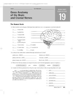
1 255 Gross Anatomy of the Brain and Cranial NervesEXErCISE 14 ObjECtIvES List the elements of the central and peripheral divisions of the nervous system. Discuss the difference between the sensory and motor portions of the nervous system, and name the two divisions of the motor portion. Recognize the terms that describe the development of the human Brain , and discuss the relationships between the terms. As directed by your instructor, identify the bold terms associated with the cerebral hemispheres, diencephalon, Brain stem, and cerebellum on a dissected human Brain , Brain model, or appropriate image, and state their functions.
2 State the differences among gyri, fissures, and sulci. Describe the composition of gray matter and white matter in the nervous system. Name and describe the three meninges that cover the Brain , state their functions, and locate the falx cerebri, falx cerebelli, and tentorium cerebelli. Discuss the formation, circulation, and drainage of cerebrospinal fluid. Identify the Cranial Nerves by number and name on a model or image, stating the origin and function of each. Identify at least four key anatomical differences between the human Brain and sheep QuIz 1. Circle the correct underlined term. The central nervous system / peripheral nervous system consists of the Brain and spinal cord.
3 2. Circle the correct underlined term. The most superior portion of the Brain includes the cerebral hemispheres / Brain stem. 3. Circle True or False. Deep grooves within the cerebral hemispheres are known as gyri. 4. On the ventral surface of the Brain , you can observe the optic Nerves and chiasma, the pituitary gland, and the mammillary bodies. These externally visible structures form the floor of the: a. Brain stem c. frontal lobe b. diencephalon d. occipital lobe 5. Circle the correct underlined term. The inferior region of the Brain stem, the medulla oblongata / cerebellum houses many vital autonomic centers involved in the control of heart rate, respiratory rhythm, and blood pressure.
4 6. Directly under the occipital lobe of the cerebrum is a large cauliflower-like structure known as the: a. Brain stem b. cerebellum c. diencephalonMatErIaLS Human Brain model (dissectible) Preserved human Brain (if available) Three-dimensional model of ventricles Frontally sectioned or cross-sectioned human Brain slice (if available) Materials as needed for Cranial nerve testing (see Table ): aromatic oils ( , vanilla and cloves); eye chart; ophthalmoscope; penlight; safety pin; blunt probe (hot and cold); cotton; solutions of sugar, salt, vinegar, and quinine; ammonia.
5 Tuning fork, and tongue depressor Preserved sheep Brain (meninges and Cranial Nerves intact) Dissecting instruments and tray Disposable glovesText continues on next 25511/13/15 11:06 AM256 Exercise 14 When viewed alongside all Earth s animals, humans are indeed unique, and the key to our uniqueness is found in the Brain . Each of us is a reflection of our Brain s experience. If all past sensory input could mysteri-ously and suddenly be erased, we would be unable to walk, talk, or communicate in any manner. Spontaneous movement would occur, as in a fetus, but no voluntary integrated func-tion of any type would be possible.
6 Clearly we would cease to be the same convenience, the nervous system, is considered in terms of two principal divisions: the central nervous system and the peripheral nervous system. The central nervous system (CNS) consists of the Brain and spinal cord, which primarily interpret incoming sensory informa-tion and issue instructions based on that information and on past experience. The peripheral nervous system (PNS) consists of the Cranial and spinal Nerves , ganglia, and sen-sory receptors. The PNS has two major subdivisions: the sensory portion, which consists of nerve fibers that conduct impulses from sensory receptors toward the CNS, and the motor portion, which contains nerve fibers that conduct impulses away from the CNS.
7 The motor arm, in turn, consists of the somatic division (sometimes called the voluntary system), which controls the skeletal muscles, and the other subdivi-sion, the autonomic nervous system (ANS), which controls smooth and cardiac muscles and glands. This exercise focuses on the Brain (CNS) and Cranial Nerves (PNS) because of their close anatomical Human BrainDuring embryonic development of all vertebrates, the CNS first makes its appearance as a simple tubelike structure, the neural tube, that extends down the dorsal median plane. By the fourth week, the human Brain begins to form as an expan-sion of the anterior or rostral end of the neural tube (the end toward the head).
8 Shortly thereafter, constrictions appear, dividing the developing Brain into three major regions forebrain, midbrain, and hindbrain (Figure ). The remainder of the neural tube becomes the spinal central canal of the neural tube, which remains continuous throughout the Brain and cord, enlarges in four regions of the Brain , forming chambers called ventricles (see Figure and b, page 265). 7. Circle the correct underlined term. The outer cortex of the Brain contains the cell bodies of cerebral neurons and is known as white matter / gray matter. 8. The Brain and spinal cord are covered and protected by three connective tissue layers called: a.
9 Lobes c. sulci b. meninges d. ventricles 9. Circle True or False. Cerebrospinal fluid is produced by the frontal lobe of the cerebrum and is unlike any other body How many pairs of Cranial Nerves are there? _____ Activity 1 Identifying External Brain StructuresIdentify external Brain structures using the figures cited. Also use a model of the human Brain and other learning aids as they are , the Brain is studied in terms of four major regions: the cerebral hemispheres, diencephalon, Brain stem, and cerebellum. Cerebral HemispheresThe cerebral hemispheres are the most superior portion of the Brain (Figure ).
10 Their entire surface is thrown into ele-vated ridges of tissue called gyri that are separated by shallow grooves called sulci or deeper grooves called fissures. Many of the fissures and gyri are important anatomical cerebral hemispheres are divided by a single deep fissure, the longitudinal fissure. The central sulcus divides the frontal lobe from the parietal lobe, and the lateral sulcus separates the temporal lobe from the parietal lobe. The parieto-occipital sulcus on the medial surface of each hemisphere divides the occipital lobe from the parietal lobe. The sulcus is not visible externally.