Transcription of Guidelines for Noninvasive Vascular Laboratory …
1 Guidelines for Noninvasive VascularLaboratory Testing: A Report from theAmerican society of Echocardiography andthe society of Vascular Medicine and BiologyRepresented by Marie Gerhard-Herman, MD, MMSc, Julius M. Gardin, MD, FASE,Michael Jaff, DO, Emile Mohler, MD, Mary Roman, MD, andTasneem Z. Naqvi, MD, FASE, RVTEXECUTIVE SUMMARYA ccompanying the rapid growth of interest inpercutaneous Vascular interventions, there hasbeen increasing interest among cardiologists inperforming Noninvasive Vascular testing using ul-trasound. In an attempt to provide recommenda-tions on the best practices in Vascular laboratorytesting, this report has been prepared by a writinggroup from the american society of Echocardiog-raphy (ASE) and the society of Vascular Medicineand Biology.
2 The document summarizes principlesintegral to Vascular duplex ultrasound includingcolor Doppler, spectral Doppler waveform analy-sis, power Doppler, and the use of indications and interpretation of ca-rotid artery, renal artery, abdominal aorta, andperipheral artery ultrasound imaging are de-scribed. A dedicated section summarizes noninva-sive techniques for physiologic Vascular testing ofthe lower extremity arteries including measure-ment of segmental pressures and pulse volumeplethysmography. The use of exercise testing inthe evaluation of peripheral artery disease, ultra-sound evaluation of the lower extremities afterpercutaneous revascularization, and the diagnosisand management of iatrogenic pseudoaneurysm(PSA) is also discussed.
3 A section on the importanttopic of Vascular Laboratory accreditation is in-cluded. Finally, additional details regarding propertechnique for performance of the various vasculartests and procedures are included in the has been increasing demand for vascularultrasound training among cardiologists in practiceand in training. For example, the recent documenton training for cardiology fellows, COCATS-2, hasrecommended 2 months of dedicated or aggregate instruction in the Noninvasive Laboratory for Level1 training in Vascular article willreview general principles, indications, and interpre-tation of Noninvasive Vascular testing of the carotidarteries, renal arteries, abdominal aorta, and periph-eral arteries.
4 Additional details regarding the tech-niques of performing Vascular ultrasound are pro-vided in the Appendix. Another article by thisworking group, Clinical Application of NoninvasiveVascular Ultrasound in Cardiovascular Risk Stratifi-cation, will review the application of carotid artery(intimal-medial thickness) and brachial artery (flow-mediated dilatation) measurements for cardiovascu-lar risk : GENERALCONSIDERATIONSV ascular testing includes duplex ultrasound andphysiologic evaluation. Vascular ultrasound testsrequire a machine equipped with 5- to 12-MHzlinear-array transducers (for the neck and extrem-ities) and to curved linear- orphased-array transducers (for the abdomen).
5 Avascular software package is required in additionto the appropriate transducers. Duplex scanningrefers to an ultrasound scanning procedure re-cording both gray scale and Doppler Brigham and Women s Hospital, Boston, MA ( ); Hospital and Medical Center, Detroit, MI ( ); Massa-chusetts General Hospital, Boston, MA ( ); University ofPennsylvania Health System, Philadelphia, PA ( ); Weill Med-ical College of Cornell University, New York, NY ( ); CedarsSinai Medical Center, Los Angeles, CA ( ). Copyright 2005 american society of Echocardiography (ASE).Property of the ASE.
6 Reprint of these documents, beyond singleuse, is prohibited without prior written authorization of the requests: The american society of Echocardiography,1500 Sunday Dr, Suite 102, Raleigh, NC 27607. (919) Am Soc Echocardiogr 2006;19 $ 2006 by the american society of includes 2-dimensional structure and motion,Doppler spectrum analysis, and color flow veloc-ity mapping. Carotid arteries, renal arteries, ab-dominal aorta, and peripheral arteries can beappropriately evaluated using this testing includes segmental pulse vol-ume recording and segmental pressure measure-ments with cuffs appropriately sized for the lowerextremities and a plethysmographic recording : PRINCIPLES APPLICABLE TO ALLVASCULAR TESTINGThe ultrasound beam is directed perpendicular tothe surface of interest to obtain the brightest echowith gray-scale imaging and optimal imaging ofthe artery wall.
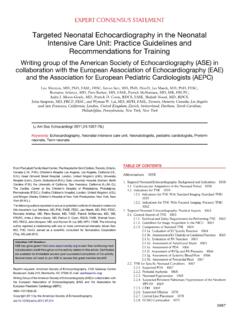
7 The perpendicular angle is oftenreadily obtained, as arteries generally are parallelto the surface of the transducer (Figure 1). For theDoppler component of duplex imaging, an angleof 60 degrees between the Doppler insonationbeam and the vessel wall should be Doppler angle becomes an important consid-eration when the velocity data are used to above 60 degrees can result insignificant overestimation of the velocity andshould be avoided. Angles that are not relevant tothe vessel wall may misrepresent the true peakvelocity3(Figure 2).Color DopplerThe pulse repetition frequency scale determinesthe degree of color saturation and is adjusted sothat normal laminar flow appears as a region ofhomogeneous color.
8 Stenosis results in the pro-duction of a high velocity jet and an abruptchange in the color flow pattern. This is identifiedas either aliasing or desaturation (whitening) ofthe color display at the site of luminal occurs when the flow velocity exceedsthe Nyquist limit and results in color display of thereverse flow direction (wrap around). The post-stenotic region demonstrates a mosaic patternindicating turbulent flow (Figure 3). Gray-scalesettings are adjusted to optimize visualization ofintraluminal plaque or thrombus at these sites ofabnormal flow. Color Doppler provides additionalinformation used to detect a significant aliasing, persistence, and bruit all indicateflow disturbance.
9 Color persistence is a continu-ous flow signal that is color of the forwarddirection only, in contrast to the alternating colorin normal persistence correspondsto the monophasic spectral Doppler waveform ofsevere stenosis. A color bruit in the surroundingsoft tissue also indicates flow disturbance. Thiscolor artifact is attributed to vibration in thesurrounding soft tissue in the presence of a highvelocity jet. Abnormalities of color flow indicatepossible stenosis that is then characterized usingpulsed wave Doppler determination of 1 Vessel of interest should be perpendicular to ultrasound beam for B-mode imaging to obtainmost distinct , Carotid B-mode image obtained with ultrasound beam perpendicular to vesselwall (arrow) demonstrates trilaminar structure of arterial , Wall structure is poorly defined withnonperpendicular angle (arrow).
10 Journal of the american society of Echocardiography956 Gerhard-Herman et alAugust 2006 Spectral Doppler Waveform AnalysisA normal pulsed wave Doppler waveform is asharply defined tracing with a narrow Dopplerspectrum indicating that blood cells are moving atsimilar speed throughout the cardiac cycle (Fig-ure 4). Flow becomes turbulent at bifurcationsand luminal narrowings causing spectral broaden-ing of Doppler waveform, with filling in of the lowvelocity region in the spectral waveform as theblood cells move at a wide range of velocities(Figure 4). The normal peripheral artery wave-form is triphasic (Figure 4).