Transcription of Hematoma volumes of spontaneous intracerebral …
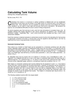
1 540 ARTICLEDOI: volumes of spontaneous intracerebral hemorrhage: the ellipse (ABC/2) method yielded volumes smaller than those measured using the planimetric methodVolumes de hematomas em hemorragias intracerebrais espont neas: o m todo da elipse (ABC/2) produziu volumes inferiores do que aqueles determinados pelo m todo planim tricoAdriano Keijiro Maeda1, Luiz Roberto Aguiar2, Carolina Martins3, Gerson Linck Bichinho4, Munir Ant nio Gariba4 Cerebrovascular accidents (CVA) include ischemic cerebral attacks, subarachnoid hemorrhage, and spontaneous intracere-bral hemorrhage (ICH). The latter is the deadliest type of CVA, accounting for 10% of total strokes1,2. Stroke is considered the third single cause of mortality in the United States of America, following cardiovascular diseases and neoplasms. In 2005, stroke was the cause of one in every 15 deaths in the USA, and 8 to 15% of patients with acute ischemic strokes and 37 to 38% of the ce-rebral hemorrhage ones died within 30 days3.
2 In addition, only 20% of patients with ICH recovered functional independence, suggesting a poor prognosis for hypertension is considered the main reason for surgical indication in subjects with ICH, especially in cases in which there is a progressive neurological deterioration5,6. The 1MD, Postgraduate Health Technology Program, Pontifical Catholic University of Paran , Curitiba PR, Brazil;2MD, MsC, PhD, Postgraduate Health Technology Program, Pontifical Catholic University of Paran ; Neurosurgeon at Hospital Santa Cruz, Curitiba PR, Brazil;3MD, PhD, Neurosurgeon at Hospital Pel pidas Silveira, Instituto de Medicina Integral Professor Fernando Figueira (IMIP), Recife PE, Brazil;4 MSc, PhD, Postgraduate Health Technology Program, Pontifical Catholic University of Paran , Curitiba PR, : Luiz Roberto Aguiar; Rua Imaculada Concei o ; 80215-901 Curitiba PR - Brasil; E-mail: of interest: There is no conflict of interest to 26 March 2012; Received in final form 26 March 2013; Accepted 02 April : To compare two different methods for measuring intracerebral hemorrhage (ICH) volume : the ellipse volume (called ABC/2), and the software-aided planimetric.
3 Methods: Four observers evaluated 20 brain computed tomography (CT) scans with spontaneous ICH. Each professional measured the volume using the ABC/2 and the planimetric methods. The average volumes were obtained, and the intra- and in-ter-rater variability was determined. Results: There is an absolute cm3 average difference between both methodologies. volumes yielded by the ABC/2 method were as much as smaller than by the planimetric one. An intra-observer variability rate of was found for the planimetric method and for the ABC/2. The inter-observer rates were and respectively. Conclusions: Both methods are reproducible. The ABC/2 yielded hemorrhage volumes as much as smaller than those measured using the planimetric methodology. Key words: cerebral hemorrhage, tomography, evaluation : Comparar dois m todos diferentes para determinar o volume da hemorragia intracerebral : volume da elipse (chamado ABC/2), e m todo planim trico auxiliado por computador.
4 M todos: Quatro diferentes observadores avaliaram as imagens de 20 tomografias cerebrais com diagn stico de hemorragia intracerebral espont nea. Cada profissional determinou o volume da hemorragia usando os dois m todos. Foram comparadas as m dias dos volumes obtidos, bem como suas variabilidades intra e interobservadores. Resultados: Foi observada dife-ren a estatisticamente significativa entre os volumes calculados por meio dos dois m todos, com uma varia o m dia absoluta de 2,24 cm3 e com volumes at 14,9% menores para o m todo ABC/2. A m dia da variabilidade intraobservador foi de 0,46% para o m todo planim trico e 0,18% para o ABC/2. As taxas de variabilidade interobservador foram de 1,69 e de 1,11%, respectivamente. Conclus es: Ambos os m todos s o reprodut veis. O volume determinado pelo ABC/2 pode ser at 14,9% menor que aquele determinado pelo m todo planim : hemorragia cerebral, tomografia, estudos de avalia Keijiro Maeda et al. intracerebral hemorrhage: volume measurementhemorrhage size, ventricular extension and expansion of the he-matoma, along with altered level of consciousness, are decisive factors in the evolution of patients7,8.
5 An increase in the ICH vol-ume is a major contributor to poor prognosis, especially when it occurs early on, since it indicates neurological deterioration9-14. Given the importance of measuring accurately the hemor-rhage lesion volume in properly determining patient prognosis, the aim of this study was to compare two methods that are cur-rently used for determining spontaneous hemorrhage volume : the ellipsoid (or ABC/2), and the was a retrospective study, carried out from October 2007 to January 2008, approved by the Ethics and Research Committee of the Pontifical Catholic University of Paran , in Curitiba, Paran , Hematoma volumes of spontaneous ICH, using patients in the acute phase, were measured by two different methods (ABC/2 and the planimetric method) of computed tomography (CT) scans, obtained by volumetric acquisition in a Somaton equipment (Siemens, Erlangen, Germany). The field of view (FOV) was selected at the time of examination according to pa-tient s characteristics (weight and position) with a pitch of 1:1 and 3 mm thick cuts.
6 Further reconstructions to 1 mm were made to all independent raters evaluated the CT scans of 20 pa-tients with spontaneous ICH, including five cases of posterior fossa and 15 of supratentorial hemorrhage. Bleeding was ob-served in 83 CT slices. Two measurements were made for each method (ABC/2 and planimetric) in two independent sessions, with at least a two-day interval. volume measurements using the ABC/2 method were performed using DicomViewer software (DicomWorks, Limoges, France). The images were transferred as DICOM files, and the tomography slices showing the hemor-rhage were individually assessed. The slice with the largest area of Hematoma was chosen for the measurements. Measure A cor-responds to the greatest diameter of hemorrhage; B to the larg-est perpendicular diameter to A; whereas C was the sum of the thickness of slices containing the hemorrhage (Fig 1A). For the sum of the slice thickness (C), one was excluded if the Hematoma area was smaller than 25% of the larger obtained place.
7 Half of the slice thickness was taking into consideration if the measured area were between 25 to 75% when compared to the largest one, as proposed by Kothari et Values for A, B, and C were then multiplied, and the final result was divided by two, which was expressed in cm3. Each observer performed two independent measurements using the ABC/2 method, with at least a two-day planimetric measurements were performed using the planning station of BrainLab neuronavigation equip-ment. The DICOM files of the CT images were transferred to the workstation using PatXFer and exported to Iplan Cranial software (BrainLab, Munich, Germany), a com-ponent of the utilities package used for planning navigation. Hemorrhage edges were individually determined for each slice using the available software tools (brush and smart-brush), as seen in Fig 1B. This software calculates the total hemorrhage volume , expressed in cm3.
8 The volumes obtained using the two methods were compared using Student s t-test for dependent samples, and the correlation between them was estimated using Pearson s correlation coefficient and tested for its major significance. Fig 1. (A) Axial computed tomography scan slice displaying the method of linear measurement using DicomWorks software; (B) definition of the Hematoma borders using Iplan (BrainLab, M nchen).542 Arq Neuropsiquiatr 2013;71(8):540-544 The method error was determined, and the results were compared for inter- and intraobserver measurements using the variance model. The intraobserver error was determined by averaging the values obtained from each observer using both measurement methods (ABC/2 and planimetric method). The interobserver error was calculated applying the differences be-tween the average values for every two observers, for the ABC/2 and planimetric methods. RESULTST here was a significant difference in the average vol-umes obtained using both methods, the absolute difference being cm3.
9 The average measurement obtained with ABC/2 method was lower than that obtained with the planimetric one. Table 1 and Fig 2 show average, minimum, Fig 2. Average and standard deviation for planimetric and ellipsoid 3. Pearson s correlation coefficient between planimetric and ellipsoid method (r= ; p< ).6050403020100-10 ABC/2-5 0 5 10 15 20 25 30 35 40 45 50 PLANIM ethodAverage (cm3)Standard deviation (cm3)p-value* (PLANI ABC/2) <0,001 Table 1. Planimetric and ABC/2 method comparison.*Student s t-test for paired samples; p< maximum values, which ranged from 25 to 75% of data. However, a good correlation between both methodologies can be observed as showed by Pearson s correlation coeffi-cient, in the two methods (r= ; p< ), as seen in Fig intraobserver error is listed in Table 2 for ABC/2 method and in Table 3 for the interobserver error was calculated using the differ-ences between the average values for every two observers.
10 Table 4 shows the results for ABC/2 and Table 5 for the pla-nimetric ABC/2 method provides an estimated value of the spontaneous ICH volume . Although several studies validate this method for clinical purposes16-21, it is considered to be the most effective for measuring ICH that have a rounded or elliptical shape20, as opposed to irregularly-shaped hemor-rhages, which are in fact more common among patients who take anticoagulants19. In a previous study, Gebel et used both meth-ods (ABC/2 and planimetric) to compare volumes of Observer 1 Observer 2 Observer 3 Observer 4 MeasurementMeasurementMeasurementMeasure ment12121212 Average (cm3) (cm3) .1 00 .1 7 Table 2. Average values of ABC/2 from each observer and differences between 1 Observer 2 Observer 3 Observer 4 MeasurementMeasurementMeasurementMeasure ment12121212 Average (cm3) (cm3) 3. Average values of the planigraphic method from each observer and differences between them.