Transcription of Hematometra - ISRJEM
1 HematometraIsraeli Journal of Emergency Medicine Vol. 5, No. 3 July 2005 - 13 Hematometra An unusual cause of suprapubic pain in an adult. Sam G Campbell MB BCh ,* Rebecca Dobson MD FRCPC ^ * Samuel G Campbell: Department of Emergency Medicine, Dalhousie University Queen Elizabeth II Health Sciences Centre, Canada ^ Rebecca Dobson: Department of Diagnostic Imaging, Dalhousie University Queen Elizabeth II Health Sciences Centre, Canada Abstract: Pelvic pain is a common presentation to the emergency department, and may be difficult to diagnose and manage. We describe the case of a young woman who presented to the emergency department with severe suprapubic pain due to Hematometra resulting from cervical stenosis following a cervical cone biopsy, performed four months prior to presentation.
2 This uncommon cause of pelvic pain should be considered in women of childbearing potential, especially in cases of prior cervical surgery or primary amenorroea. Investigational modalities include pelvic ultrasound, CT and MRI. MeSH Words: Hematometra , hematocolpos, pelvic pain. Introduction Pelvic pain is a common presentation to the emergency department (ED), and symptoms of the various causative syndromes overlap considerably1. Treatment options are often ineffective 2,3. Chronic pelvic pain is particularly distressing for patients, misdiagnosis and misdirected therapies are frequent5 and the cause of the pain is may be elusive2 4. Acute pelvic emergencies may present as sub-acute or chronic pelvic pain, and it is imperative that physicians maintain a high index of suspicion for conditions that require urgent treatment 1.
3 Differential diagnoses to consider in women presenting with pelvic pain are listed in table 1. Case Report A 30 yr old female presented to the Emergency Department with a history of 2 months of steadily increasing sharp and cramp like lower abdominal pain which had become significantly worse in the previous five days. She had not experienced dysuria or vaginal discharge, but had experienced significant dyspareunia in the previous month. Seven months before her ED presentation, she had undergone an uncomplicated caesarian section for failure to progress during labor after an otherwise normal first pregnancy. At that time she requested, and received an injection of depo-medroxyprogesterone acetate (DMPA) at the HematometraIsraeli Journal of Emergency Medicine Vol.
4 5, No. 3 July 2005 - 14 time of discharge for contraception. Four months prior to this emergency presentation, she underwent a cold knife cone biopsy for persistent Pap smear findings of high grade squamous intra epithelial neoplasia. The procedure went well with no complications recorded. She reported that her periods, that had been sparse since her caesarian, had stopped completely after the latter procedure. Other medical history was non-contributory. Examination showed an obese female in acute distress, crying with pain. Her lower abdomen was tender throughout, although neither percussion nor rebound tenderness was present.
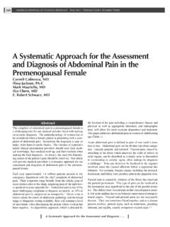
5 In spite of intravenous narcotics, pelvic examination was limited by pain and severe cervical excitation tenderness. The cervical os could not be visualised on speculum examination nor was it possible to palpate it. Clinical evaluation of her adnexae was similarly restricted by severe tenderness. High vaginal swabs were sent for gonococcus and chlamydia culture. Her urine beta-human chorionic gonadothrophin was negative, WBC X109, Hb 126 g/L platelets 289 X 109. Transvaginal ultrasound (fig 1.) showed a large uterus distended with mobile fluid, containing multiple low level echoes. The proximal cervical was also distended with fluid. No adnexal or focal uterine masses were present.
6 She was admitted to the gynecology service, where examination under general anesthetic confirmed a bulky, 14 week uterus. Visualization of the cervix demonstrated a smooth, bluish area in the center of the cervix. Attempts at dilation of the cervix with hegar dilators were unsuccessful, and the blue area was incised with a # 1 scalpel. The opening created was dilated with hegar dilators up to a size # 9 dilator. Suction curettage removed a large amount of old blood. The procedure was complicated by a 1 cm cervical laceration, that was sutured with 2/0 chromic. There was no reported growth from the culture samples taken while the patient was in the ED. She was discharged the following day with minimal symptoms after a repeat DMPA injection.
7 At 6 months follow up she had still not menstruated, but was free of pain and repeat ultrasound was normal. Discussion We present the case of a young woman presenting with Hematometra four months after a cone biopsy and 7 months after a caesarian section. The history of secondary amennorrea was clouded by the history of DMPA injection, a common cause of secondary amenorrhoea6. The consent form signed by the patient before the cone biopsy made no mention of Hematometra or cervical stenosis as possible complications. Hematometra denotes the retention of menstrual products in the uterine cavity. Obstruction of the female genital outflow tract is ,8 The commonest of these syndromes is hematocolpos (accumulation of menstrual blood in the vagina) or secondary hematocolpometra (accumulation in the uterus and vagina) resulting from congenital malformation, most commonly Table 1.
8 Differential diagnoses of pelvic pain in the ED Pregnancy related: - Ectopic pregnancy - Miscarriage (threatened/inevitable) - Septic abortion - Premature labor - Abruption placentae - Red degeneration of a uterine fibroid - Non Pregnant:* - Pelvic inflammatory disease - Tubo-ovarian abscess - Ovarian cyst rupture/torsion - Adnexal torsion - Hematometra /hematocolpos - Endometriosis or adenomyosis - Trauma/sexual assault - Dysmenorrhoea - Mittelschmerz Non-gynecologic* - Urinary retention - Appendicitis - Incarcerated hernia - Diverticulitis - Inflammatory bowel disease - Ureteral colic - Urinary tract infection - Somatoform disorder - Musculo-skeletal pain *may also occur in pregnant patients.
9 HematometraIsraeli Journal of Emergency Medicine Vol. 5, No. 3 July 2005 - 15 Figure 1 :Saggital transvaginal ultrasound of retroverted uterus and cervix distended with fluid containing mobile low level echoes (cervix on the left, fundus on the right) imperforate hymen. Diagnosis is usually made in childhood, or at menarche with the advent of cyclical pain and primary Uncommon presentations at this age include urinary retention10,11, recurrent urinary tract infection, and back The condition has been misdiagnosed as appendicitis, when the blood-filled fallopian tube (hematosalpinx) leaks into the abdominal ,17 Hematocolpos has also been described in elderly women following vaginal occlusion secondary to radiotherapy, vaginal fibroma, and labial synechiae following infection or inflammatory Hematometra following cone biopsy is a recognized, but rare Luesley et al.
10 Described of 915 patients presenting with this condition, and noted an association between the incidence of cervical stenosis and the length of cone removed at the time of The condition usually only manifests some months to years after the procedure; Claman & Lee, in a series of 1008 cone biopsies collected over 10 years, described no cases of Hematometra , at 6 week Physical examination may reveal a suprapubic mass, which may be further palpated on rectal or vaginal examination. The uterine cervical os, may appear normal, invisible, or may show a dimple24, or a blue bulging membrane. Pelvic ultrasound is considered a first line imaging procedure21 although it may have limitations differentiating a blood filled uterine cavity from an inflammatory Endometriosis and molar pregnancy would be included in the ultrasound differential diagnosis.