Transcription of Histology Specimen Collection - Iowa Path
1 Histology Specimen Collection Histology Specimens RECOMMENDATIONS FOR ANATOMIC PATHOLOGIC TISSUE Collection . The following instructions will help us provide you with the best turn around time and most accurate pathology reports. 1. Fixation A. Tissue Specimen should be placed in a container with 10% formalin (formaldehyde) immediately after removal to prevent drying and autolysis artifact. Fasten the lid securely to prevent leakage in transit. B. A Specimen in formalin can be kept 1-2 days before processing. No refrigeration is needed. C. Large masses fix optimally if incised or sectioned, although this might compromise assessment of margins. Avoid oversectioning or mutilation. Masses within hollow organs/cysts fix better if exposed to fixative. EXAMPLE: Bowel segments should be opened opposite the surface of tumor. 2. Identification A.
2 Write the patient's name and tissue site on each container label. The chart number instead of the patient's name is unacceptable. The physician's name is not necessary. If a stamper plate is used, please write the site on that label. Do not write patient information on the lid of the container. B. Only one completed request form is necessary for all tissue specimens collected during the same procedure. If more tissue is subsequently submitted from the patient ( following a frozen section on the same case), please indicate on the requisition that there was tissue previously submitted. C. It is important to print or write the patient's first and last name on the tissue request form. Use the patient's formal name if the patient goes by a nickname. Also include the patient's date of birth, sex and day of surgery. D. Complete the patient history and clinical findings in the provided space.
3 Note previous biopsies done on that site. Also, note if there are correlating pap smears or cultures. Identify suture markers. E. Any special requests should be noted on the request form ( "Please call results ASAP). Document if assessment of margins/adequacy of excision is important. F. Fill out insurance information, including Medicare, patient's address and phone number. G. Two similar specimens in one container ( vas deferens) can be identified by attaching a suture to one Specimen . Document on the request form which Specimen the suture indicates. H. Place the formalin containers (one case only) in the main compartment of the biohazard Specimen transport bag. Fold the tissue requisition and place in the side pocket. I. Alert the courier to special storage of frozen or refrigerated specimens. 3. Recommended Container Sizes Iowa Pathology Associates offers several container sizes for Specimen Collection .
4 It is recommended that an appropriate size of container be used for Specimen fixation. An adequate volume of fixative or a ratio of 20:1 is used in a container of an appropriate size. This avoids distortion of fresh tissue and ensures good quality fixation. Some suggested container size for Specimen Collection : 20cc pre-filled formalin containers small biopsies, skins, needle biopsies, small specimens 120 cc pre-filled formalin containers appendix, gallbladder, large biopsies, lipomas Small tubs appendix, gallbladder, small uterus, small ovarian cysts, large lipomas Medium tubs uterus, small colon resection, small placenta, small mastectomy Large tubs larger placenta, large colon, large breast. 4. Tissue Culture A. For optimum results place the tissue in a sterile urine container filled with sterile saline. Refrigerate until courier pickup.
5 Fill out a laboratory request form for the culture. If the same tissue is also submitted for routine Histology , complete a tissue request form and put in the same transport pouch. Indicate that the tissue is also being submitted for culture on the tissue request form. B. For toenail clippings, collect in a sterile urine cup with no fixative. Specify if a culture is needed from the same Specimen container on the requisition. Submit two request forms one for the culture and one for the Histology processing. 5. Gynecology Specimens A. If the Specimen is a cervical cone, the Specimen should be opened, spread out, and pinned on a paraffin disk provided by our facility. Pins are not provided. Place the tissue face down in a container pre-filled with formalin, using enough formalin to cover the disk. If clinically significant, indicate on the request form if the cone has been opened at a certain site ( 12:00).
6 B. Products of Conception tissue passed by the patient at home should be placed in a glass or plastic container with a lid and refrigerated. As soon as the Specimen is received at your location, transfer the Specimen to a formalin filled container. Document time of at home Collection and time transferred to formalin on the requisition. C. Choose villi and fetal parts for chromosome analysis study. Place in a sterile urine cup and cover with sterile saline or RPMI transport medium. Refrigerate until courier pickup. Do not freeze. Indicate on the tissue request form that a chromosome analysis study is wanted and the Collection medium used. 6. Skin Specimens A. A margin can be identified with a suture. Tie the suture loosely for easy removal. B. If clinically significant, the Specimen should be oriented with a suture to identify one landmark "12:00".
7 Indicate sutured apex or margin on the request form. C. For cases involving re-excision include patient history, the previous surgical number and diagnosis. D. Tissue being submitted for direct immunofluorescent studies (IF or DIF), requires being put into Zeus transport media immediately after Collection . To order vials containing Zeus transport media call 515-241- 8860. 7. Lymph Nodes Contact our medial secretaries at 515-241-8867 at least 24 hours in advance if a pathologist is to be present for Collection of the Specimen . A. Culture studies - Obtain a sterile 2 x 3 mm lymph node sample. Submit in a sterile urine cup. B. Touch Preps It is recommended that a lymph node be bisected (or sliced transversely at 3 mm intervals). Gently touch the cut surface to four to six clean microscopic slides. Air dry the slides and place in a slide carrier.
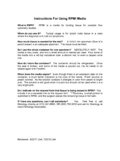
8 Submit in a separate transport bag to avoid the contact of formalin fumes. C. Immunophenotypic Studies If lymphoma is a diagnostic concern, one 2- 3 mm slice of node should be placed in RPMI. RPMI is a transport media for flow cytometry studies and can be ordered by calling the Histology laboratory (515-241-8860). The media comes in a stock bottle and only a small amount needs to be transferred into a red top tube, a dilution vial or even a sterile capped urine cup. The Specimen should be refrigerated until the courier arrives for pick-up. Call the Histology laboratory (515-241- 8860) to notify the staff that an RPMI Specimen is being sent. D. Note on request form if the tissue is also submitted in RPMI. E. Place the remaining tissue in formalin. 8. Frozen Section Evaluation Contact our medical secretaries at least 24 hours (515-241-8866) in advance to confirm scheduling.
9 If there is a delay between removal of tissue and frozen section, place tissue in saline moistened gauze to prevent desiccation. Do not place tissue in formalin. For specimens requiring needle radiographic localization, availability of mammographic services should be coordinated with frozen section scheduling. 9. Bone Marrow Aspirate and Biopsy Contact our medical secretaries at least 24 hours (515-241-8866) in advance if the pathologist is to perform the bone marrow procedure. A. Biopsy Specimen : If a biopsy Specimen is obtained, place in 10%. formalin. B. Aspirated Specimen : Place of the aspirate material in a small EDTA. tube (Lavender top). Invert tube several times. Pour remaining of aspirate into a petri dish. Prepare 8-12 crush prep slides by placing one drip of unclotted aspirate containing visible marrow particles on each of 4- 6 slides.
10 Take a second slide and put on top of the first slide and gently press the two slides together to "crush" the particles. Then slide or pull the two slides apart horizontally. Label the slide with patient name, date, and Specimen types, bone marrow aspirate. The remaining aspirate Specimen can be placed in 10% formalin, after clotting. C. If an aspirate Specimen cannot be obtained or particles are not observed in the aspirate Specimen , make four touch prep slides with the biopsy Specimen before placing it into formalin. This is done by gently pressing a glass slide against the biopsy. D. Air dry both the aspirate slides and touch prep slides. Place all slides in a slide carrier. Submit slides and formalin specimens in separate transport bags to protect the slides from formalin fumes. E. If flow cytometry, cytogenetics, or special Specimen handling is required, call the lab (515-241-8860) for special instructions.


