Transcription of How Not to Miss a Bicuspid Aortic Valve in the ...
1 ECHO ROUNDS Section Editor: E. Kenneth Kerut, Not to Miss a Bicuspid Aortic Valve in theEchocardiography LaboratorySalvatore J. Tirrito, and Edmund Kenneth Kerut, , Section of Cardiology, Tulane University Health Sciences Center, New Orleans, Louisiana Heart Clinic of Louisiana, Marrero, Louisianabicuspid Aortic Valve ,echocardiography,congenital heart diseaseAddressforcorrespondenceandreprin trequests:Salvatore J. Tirrito, , Tulane University Health Sci-ences Center, Section of Cardiology SL-48, 1430 Tulane Av-enue, New Orleans, LA 71112. Fax: 504 529-9649; and two-dimensional (2D) diastolic frame in the parasternal long-axis view of a Bicuspid Aortic Valve witheccentric closure (arrows). Although not diagnostic, an eccentric line of closure should prompt one to evaluate the Aortic valveclosely for BAV. LA=left atrium. (Modified with permission from: Kerut EK, McIlwain, Plotnick: Handbook of Echo-DopplerInterpretation 2nd Ed, Blackwell Publishing, Elmsford, New York, 2004, p.)
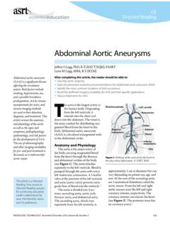
2 84)Diagnosis of Bicuspid Aortic Valve (BAV)in the busy echocardiography laboratory isimportant, as it is a relatively common cardiaccongenital defect (up to 3% of the popula-tion),1and requires a recommendation forVol. 22, No. 1, 2005 ECHOCARDIOGRAPHY: A Jrnl. of CV Ultrasound & Allied AND KERUTF igure (left panel) and systolic (right panel) two-dimensional (2D) short-axis image of a BAV. Indiastole the raphe may appear as a commissure (arrow), appearing to be a normal tri-leaflet Valve . However, thesystolic frame has a typical fish-mouth appearance. (With permission from: Kerut EK, McIlwain, Plotnick:Handbook of Echo-Doppler Interpretation 2nd Ed, Blackwell Publishing, Elmsford, New York, 2004, p. 84)endocarditis oc-curs in isolation but is associated with otheranomalies in 20% of cases. The most commonassociated anomaly is coarctation of the aorta,but occasionally patent ductus arteriosus(PDA), ascending Aortic aneurysm, and De-Bakey Type I Aortic dissection is 5 One-half of patients younger than 75 yearsold, with symptomatic Aortic stenosis, stenosis appears to progressmost rapidly when the leaflets are unequalin size and have an anteroposterior leafletconfiguration.
3 Severe Aortic regurgitation (AR)is found in 3% of patients with there exists familial clustering of BAV(male: female ratio of 3:1), echocardiographicscreening of first-degree family members ,9 ABAVmay have a right and left cusp (the ori-gin of the right coronary artery is in the rightcusp, and left coronary artery in the left cusp),or an anterior and posterior cusp (both rightand left coronary arteries originate in the an-terior cusp). M-mode of BAV may demonstratean eccentric line of closure (Fig. 1) but an ec-centric line of closure may also be seen in pa-tients with a tricuspid leaflet Aortic Valve , anda normal line of closure may be seen in pa-tients with BAV. From the parasternal shortaxis, Aortic Valve anatomy may appear normalin diastole, as a raphe may simulate three com-missural lines (Fig. 2). However, in systole thevalve will have a fish-mouth appearance. Inthe parasternal long axis, the Valve may ap-pear domed in systole (Fig.)
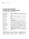
4 3) and prolapse indiastole. With BAV, the ascending aorta mayhave a normal appearance; however it should beFigure parasternal long-axis view of a patientwith a BAV. Doming (arrow) is noted. Normally the leafletshould be parallel to the Aortic wall when fully open duringsystole. LA=left atrium; RV=right : A Jrnl. of CV Ultrasound & Allied 22, No. 1, 2005 Bicuspid Aortic Valve inspected for Aortic root size and structure, asascending Aortic aneurysm and dissection maybe pointers to look for a BAV include:(i) From a parasternal long-axis window, lookfor systolic Valve doming (normally theleaflets are parallel to the aorta), and di-astolic Aortic Valve prolapse.(ii) As Aortic regurgitation (AR) is not normal, any AR noted by color Doppler warrants acloser look at the Aortic Valve , to make sureit is not Bicuspid .(iii) As a matter of routine, one should alwaysimage the descending aorta from thesuprasternal window, particularly with , Aortic coarctation, or PDA shouldprompt the sonographer to closely evaluatethe Aortic Valve .
5 (iv) As a confirmatory step in our laboratory, wehave a checkbox on our echo worksheet,for the sonographer to identify the aorticvalve as either tricuspid, Bicuspid , or can-not tell (difficult image). This is done whileimaging in systole and diastole from aparasternal short-axis view.(v) With a tricuspid leaflet Aortic Valve , oneshould note the Valve commissures extend-ing to the base of the Valve . If this cannotbe clearly visualized, consider Sabet HYBA, Edwards W, Tazelaar H, et al: Con-genitally Bicuspid Aortic valves : A surgical pathologystudy of 542 cases (1991 through 1996) and a litera-ture review of 2,715 additional Clin Proc1999;74:14 Grant RT, Wood JE, Jones TD: Heart Valve irregu-larities in relation to sub-acute bacterial ;14:247 Roberts WC: The congenitally Bicuspid Aortic Valve : Astudy of 85 autopsy J ;26 Roberts CS, Roberts WC: Dissection of the aorta as-sociated with congenital malformation of the Cardiol1991;17(3):712 Nistri S, Sorbo MD, Marin M, et al: Aortic root dilata-tion in young men with normally functioning bicuspidaortic ;82(1):19 Pomerance A: Pathogenesis of Aortic stenosis and itsrelation to Heart.
6 34:569 Guiney TE, Davies MJ, Parker DJ, et al: The aetiologyand course of isolated severe Aortic regurgitation: Aclinical, pathological, and echocardiographic ;58:358 Campbell M: Calcific Aortic stenosis and congenital bi-cuspid Aortic Heart ;30 Huntington K, Hunter AG, Chan KL: A prospectivestudy to assess the frequency of familial clustering ofcongenital Bicuspid Aortic Cardiol1997 Dec;30(7):1809 22, No. 1, 2005 ECHOCARDIOGRAPHY: A Jrnl. of CV Ultrasound & Allied