Transcription of How to Interpret Noninvasive Vascular Testing and Diagnose ...
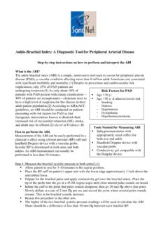
1 How to Interpret Noninvasive Vascular Testing and Diagnose peripheral Vascular disease David Campbell, MA FRCS Surgeon, Beth Israel Deaconess Medical CenterAssociate Professor of SurgeryHarvard Medical School Clinical Diagnosis Claudication versus Spinal Stenosis Ischemic Rest Pain versus Neuropathic Pain Location of foot lesions ischemic versus neuropathic Absence of symptoms does not rule out significant ischemiaSigns of PVD Pulse examination. Frequently inaccurate due to calcified vessels. Inflow versus outflow disease Autonomic neuropathy Dependent RuborNon Invasive Studies in PVD Many sophisticated tests available eg Ankle Brachial Indices, Segmental pulse volume recordings, Duplex ultrasound, Transcutaneous oxygen, Xenon flow studies. Most useful and cost effective is a hand held Doppler to assess wave formHand Held Doppler~~Interpreting the Ankle Brachial IndexAdapted from Hirsch AT.
2 Family Practice Recertification. 2000;22 > vesselsIDENTIFICATION WITH INDIRECT Testing CAPABILITY INDIRECT Testing COMPONENTS : Reliable & InexpensiveABI (Ankle Brachial Index)Multiple Level Segmental Pressures Using Doppler / Pneumatic CuffsMultiple / Single Level Pulse Volume Plethsymography (PVR)Digital Pressures / Plesthythmography (PPG)TBI (Toe Brachial Index) or DBI (Digital Brachial Index)Maneuver MeasurementsTransthoracic Outlet ExaminationCold Immersion TestingINDIRECT TESTINGIDENTIFICATION WITH INDIRECT Testing CAPABILITY INDIRECT Testing : ABISome Considerations : False Elevation Of Values / ABI:Arterial Wall (Medial) Calcification : Common In Diabetics / Renal Failure Pt s, Chronic AnticoagulationIndex Usually / Greater Than 250-300 mmHgUse Toe Pressure(s) More True Vascular Status If False Elevation SuspectedDoes Not Affect Doppler / PVR MeasurementsINDIRECT TESTINGIDENTIFICATION WITH INDIRECT Testing CAPABILITY INDIRECT Testing : ABIFALSE ELEVATION AT.
3 84 TOE Index RevealingINDIRECT TESTINGIDENTIFICATION WITH INDIRECT Testing CAPABILITY INDIRECT Testing : ABIV ariable Criteria #1 ABI = .9 Patients With Borderline Or Normal Resting ValuesCompare Pre / Post Exercise ValuesABI = .6 .9 Suspected Claudication SymptomsCompare Pre / Post Exercise ValuesABI = < .5 Exercise Testing Not NecessaryMost Likely Rest Pain*Always Compliment ABI With Doppler Waveform Morphology*Gerlock AJ, Gianani VL, Krebs C: Applications of Noninvasive Vascular Techniques, Philadelphia, Saunders, 1988 INDIRECT TESTINGINDIRECT Testing IDENTIFICATION WITH INDIRECT Testing CAPABILITY INDIRECT Testing : SEGMENTAL PRESSURES Can Localize Segment / Location Of disease Vertical Pressure Comparisons Horizontal Pressure Comparisons Artifacts To Consider 4 Cuff Or 2 Cuff MethodINDIRECT Testing IDENTIFICATION WITH INDIRECT Testing CAPABILITYINDIRECT Testing : SEGMENTAL PressuresSome Common Values Stratifying disease : Levels Of DiseaseAORTOILIAC: Thigh / Brachial index.
4 8 Stenosis Thigh / Brachial index < Iliac occlusionReduced high thigh pressure may also result from combination of:CFA Occlusion / StenosisSFA occlusion / StenosisPFA Occlusion / Stenosis Applications of Noninvasive Vascular Techniques: Gelock, Guianani, Krebs;Saunders, 1988: Ch. 17, 299-322. Segmental Pressures and Doppler Velocity Waveforms in the Evaluation of peripheral Arterial Occlusive disease : C. Burnham, BSN,RN, Journal of Vascular Technology 18[5] 249-255, Testing IDENTIFICATION WITH INDIRECT Testing CAPABILITYINDIRECT Testing : SEGMENTAL PressuresSome Common Values Stratifying disease : Levels Of DiseaseSFA disease : > 30 mmHg gradient between high thigh pressure and above knee pressure. > 25 mmHg gradient between above knee pressure and contra lateral above knee pressure.
5 Applications of Noninvasive Vascular Techniques: Gelock, Guianani, Krebs;Saunders, 1988: Ch. 17, 299-322. Segmental Pressures and Doppler Velocity Waveforms in the Evaluation of peripheral Arterial Occlusive disease : C. Burnham, BSN,RN, Journal of Vascular Technology 18[5] 249-255, Testing IDENTIFICATION WITH INDIRECT Testing CAPABILITYINDIRECT Testing : SEGMENTAL PressuresSome Common Values Stratifying disease : Levels Of DiseasePOPLITEAL disease : > 30 mmHg gradient between above knee & below knee > 15 mmHg gradient between below knee & contra lateral below knee Applications of Noninvasive Vascular Techniques: Gelock, Guianani, Krebs;Saunders, 1988: Ch. 17, 299-322. Segmental Pressures and Doppler Velocity Waveforms in the Evaluation of peripheral Arterial Occlusive disease : C.
6 Burnham, BSN,RN, Journal of Vascular Technology 18[5] 249-255, Testing IDENTIFICATION WITH INDIRECT Testing CAPABILITYINDIRECT Testing : SEGMENTAL PressuresSome Common Values Stratifying disease : Levels Of DiseaseTIBIOPERONEAL disease : > 30mmHg gradient between below knee & ankle > 15 mmHg gradient between ankle pressure & contra lateral ankle Applications of Noninvasive Vascular Techniques: Gelock, Guianani, Krebs;Saunders, 1988: Ch. 17, 299-322. Segmental Pressures and Doppler Velocity Waveforms in the Evaluation of peripheral Arterial Occlusive disease : C. Burnham, BSN,RN, Journal of Vascular Technology 18[5] 249-255, Testing IDENTIFICATION WITH INDIRECT Testing CAPABILITYINDIRECT Testing : SEGMENTAL PressuresSome Common Values Stratifying disease : Levels Of DiseaseDIGITAL ARTERY disease : Digital pressure < 60% of ankle pressure Toe / Brachial index < Toe systolic pressure < 30 mmHg Indicates a probable non-healing lesion Digit pressures < 80% of the brachial pressure indicate proximal disease Applications of Noninvasive Vascular Techniques: Gelock, Guianani, Krebs;Saunders, 1988: Ch.
7 17, 299-322. Segmental Pressures and Doppler Velocity Waveforms in the Evaluation of peripheral Arterial Occlusive disease : C. Burnham, BSN,RN, Journal of Vascular Technology 18[5] 249-255, Testing IDENTIFICATION WITH INDIRECT Testing CAPABILITY INDIRECT Testing : PULSE VOLUME RECORDING / PLETHYSMOGRAPHYTO RECORD THE CURVE OF FILLING Greek OriginationINDIRECT Testing IDENTIFICATION WITH INDIRECT Testing CAPABILITY INDIRECT Testing : PULSE VOLUME RECORDING / PLETHYSMOGRAPHYWhat Does It Do ? Measures Changes In Pressure Within The Cuff Pressure Changes In The Volume Of The Cuff Or BladderRelates To Pressure Changes Within Limb Volume Detected Cuffs At Various Levels Compare Volume Changes Between Horizontal + Vertical Levels Typically Inflated To 65 mmHg (Protocols Vary)Enough To Provide Contact To Skin And To Reflect Pulsatility Amplitude Changes On The GraphINDIRECT Testing IDENTIFICATION WITH INDIRECT Testing CAPABILITY INDIRECT Testing : PULSE VOLUME RECORDING / PLETHYSMOGRAPHYPVR Influenced By : Blood PressureVolume Of Blood (Infection ?)
8 Cellulitis ?)Position Of ExtremityOverall Size Of ExtremityCardiac Stroke VolumeMay Even Be Different On Same Patient B/W VisitsLarge Habitus + Edema Will Attenuate PVR Presentation / WaveExcessive or Not Enough Cuff InflationINDIRECT Testing IDENTIFICATION WITH INDIRECT Testing CAPABILITY INDIRECT Testing : PULSE VOLUME RECORDING / PLETHYSMOGRAPHYUSEFUL FOR: Determining Level Of disease : Aorto-Iliac + Outflow Proximal SFA / DFA InvolvementMid SFA / Abductor Canal Popliteal / TibialOther Uses: Pre + Post Exercise MeasurementsIntra-Op MonitoringPost-Op EvaluationsHealing PotentialConfirmation Of Rest Pain SymptomsINDIRECT Testing IDENTIFICATION WITH INDIRECT Testing CAPABILITY INDIRECT Testing : PULSE VOLUME RECORDING / PLETHYSMOGRAPHYCONTOUR PRESENTATION.
9 NORMAL Higher Amplitude BKDicrotic Notch Present(Arterial Pulse Reverse Component)INDIRECT Testing IDENTIFICATION WITH INDIRECT Testing CAPABILITY INDIRECT Testing : PULSE VOLUME RECORDING / PLETHYSMOGRAPHYCONTOUR PRESENTATION: NORMALD icrotic Notch More Pronounced W/ Vasoconstriction Less Pronounced / DisappearsW/ VasodilationW/ Prox. ObstructionINDIRECT Testing IDENTIFICATION WITH INDIRECT Testing CAPABILITY INDIRECT Testing : PULSE VOLUME RECORDING / PLETHYSMOGRAPHYCONTOUR PRESENTATION: MILD (Criteria Varies)Loss Of Dicrotic NotchUpstroke Is Less SteepRounded PeakDown slope BowingINDIRECT Testing IDENTIFICATION WITH INDIRECT Testing CAPABILITY INDIRECT Testing : PULSE VOLUME RECORDING / PLETHYSMOGRAPHYCONTOUR PRESENTATION: (Criteria Varies)MODERATESEVERELess Amplitude With SeverityINDIRECT Testing IDENTIFICATION WITH INDIRECT Testing CAPABILITY INDIRECT Testing : PULSE VOLUME RECORDING / PLETHYSMOGRAPHYCONTOUR PRESENTATION: (Criteria Varies)Tachycardia Camouflaged NotchObviously Normal INDIRECT Testing IDENTIFICATION WITH INDIRECT Testing CAPABILITY INDIRECT Testing .
10 PULSE VOLUME RECORDING / PLETHYSMOGRAPHYCONTOUR PRESENTATION: (Criteria Varies)Popliteal / Tibial Trunk DiseaseINDIRECT Testing IDENTIFICATION WITH INDIRECT Testing CAPABILITY INDIRECT Testing : PULSE VOLUME RECORDING / PLETHYSMOGRAPHYCONTOUR PRESENTATION: (Criteria Varies)Well Developed Collaterization ?INDIRECT Testing IDENTIFICATION WITH INDIRECT Testing CAPABILITY INDIRECT Testing : CW DOPPLERIDENTIFICATION WITH INDIRECT Testing CAPABILITY INDIRECT Testing : CW DOPPLER Reflects The Compliance and Elasticity Of The Artery Triphasic Morphology Normal Loss Of Phasicity Due To Decreased Elasticity / Compliance Of The Artery As disease ProgressesReversal In Early DiastoleForward In Late DiastoleNORMALINDIRECT TESTINGIDENTIFICATION WITH INDIRECT Testing CAPABILITY INDIRECT Testing : CW DOPPLER Reflects The Compliance and Elasticity Of The Artery Loss Of Phasicity Due To Decreased Elasticity / Compliance Of The Artery As disease ProgressesForward In Late Diastole LossBIPHASICMONOPHASICINDIRECT TESTINGWhat's The Difference?