Transcription of How to prepare aceto-orcein squashes, especially …
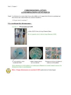
1 How to prepare aceto-orcein squashes , especially for pachytene chromosomes. Edward G. Barry and David D. Perkins Background aceto-orcein squashes of meiotic chromosomes provided the first definitive information on the karyotype and chromosome morphology of Neurospora (McClintock 1945, Singleton 1953; see Perkins 1992). Orcein staining still provides the clearest definition of chromomere morphology at pachytene (Figure 1). (For McClintock's drawings of the seven pachytene bivalents see Perkins 1992 or the frontispiece in Perkins et al. 2001.) The nucleolus is stained only lightly, allowing underlying chromosomes of the pachytene karyotype to be visualized. The fine detail revealed by this technique is superior to that obtained using haematoxylin or other procedures. The method has not been widely used, however, because it requires exceptional patience and skill. Preparations are short lived and cannot be made permanent.
2 Orcein from some sources does not give good preparations. The cytoplasm is highly vacuolated (although the nucleoplasm is clear), in contrast to preparations made with haematoxylin according to a protocol in which chloroform clears the cytoplasm. Despite these drawbacks, the method was used effectively for critical basic studies of meiosis and ascus development, meiotic behavior of chromosome rearrangements, and for assigning genetic linkage groups to cytologically defined chromosomes (Singleton 1952, Barry 1969, Perkins and Barry 1977). The following account of the method is copied (with minor modifications) from Barry (1966). Procedure The following procedure has given satisfactory preparations of meiotic chromosomes of Neurospora crassa. Good results are obtained almost routinely and occasional excellent preparations are found. Procedures are described in detail because some of our techniques are unorthodox or because it man not be known whether a particular feature contributes to good results or whether it is irrelevant.
3 Crosses and timing: Crosses are made by growing one parental strain (usually wild type) as protoperithecial parent on 15 to 20 ml of synthetic crossing medium (Westergaard and Mitchell 1947) with 2% sucrose and 2% agar in petri plates at 25 C. At 6 days the surface of the protoperitheciating culture is dusted with conidia (or scrubbed with hyphal fragments) from the fertilizing parent. To determine closely when to begin fixing material, a few perithecia may be picked from the cross beginning 3 days after fertilization, squeezed into a drop of water under a dissecting microscope, and examined for the presence of young asci. Usually, pachytene stages are first found 3 to 4 days after crossing, and condensed chromosome stages (diakinesis and later) are abundant from 4 days on. Material is usually fixed at 4, 5, and 6 days after crossing. The sample in which the desired stages are most abundant is used for observations.
4 Fixation: Strips of agar bearing perithecia are cut from the petri plates and immersed in freshly made fixative in closed vials or 10 75 mm tubes stoppered with No. 0 corks. The fixative consists of 6 parts absolute alcohol, 1 part glacial acetic acid, and 1 part 85% lactic acid. The fixed material is kept in a deep freeze or the freezer compartment of a refrigerator. Although some fixed material has given good quality preparations after being stored for 2 years, there is a suggestion of degeneration with time. For best results it is recommended that the fixed material not be stored more than 2 or 3 months. Stain: Successful staining requires that fresh stain be made each day. It is convenient to make up the stain components in bulk. The liquid components, 47 ml glacial acetic acid, 20 ml lactic acid solution (1 ml 85% lactic acid in 24 ml distilled water), 5 ml 1 N hydrochloric acid, and 28 ml distilled water are mixed 1and stored at room temperature.
5 Natural orcein is weighed out into 10 75 mm tubes in 100 mg lots. For each 100 mg batch, 5 ml of the liquid portion of the stain is mixed with 100 mg orcein in a 50 ml beaker covered by a petri plate bottom that contains two ice-cubes. The stain solution is refluxed over a low flame (alcohol lamp or Bunsen burner pilot) for 4 minutes after it begins to boil . The stain is cooled for 2 hours by placing the beaker in the melting ice-cube water in the petri bottom, and it is then filtered through #1 Whatman filter paper into a small dropping bottle. [Best results have been obtained using orcein from Edward Gurr, Ltd. London. However, that source has now gone out of business. Once existing supplies are gone, a new source must be found.] Dissection of perithecia; preparation of slides: Perithecia are taken from the fixative, scraped from the agar block, placed in a drop or two of stain on the frosted end of a carefully cleaned slide (Aloe #58952) and examined under a dissecting microscope at about 30 magnification using transmitted light from below.
6 A pair of dissecting needles is used to scrape mycelial fragments and remnants of agar away from about ten perithecia. The perithecia are then transferred to a fresh, small drop of stain near the middle of the slide. The contents of each perithecium are expressed through the ostiole into the stain, using a slightly flattened and bent or chisel-shaped steel needle (about No. 1 embroidery needle). Perithecial walls and other debris are removed. If large clumps of asci are present, they are teased or chopped apart, or even removed. The best staining is found in separated and broken asci, and these are most readily obtained when the perithecial contents squirt through the ostiole rather than when a clump of asci emerges intact from a broken perithecial wall. A ring of crystallized stain often forms during this operation, and it may cause difficulty later by holding up the coverglass. We have used two methods to avoid the problem: either the dried stain is put back into solution by a rapid mixing with the needles, or fewer perithecia are used, the work goes faster, and the smaller amount of precipitate does not interfere when the cover is lowered.
7 A coverglass is dropped over the stain and permitted to flatten the asci without other external pressure. 18 mm square No. 1 coverglasses are convenient, and are less readily broken than No. 0. When a small drop of stain has been used and no debris or dust particles are present, a thin preparation results in which some asci may even burst from the pressure when the coverglass is drawn down by surface tension as the stain spreads under it. If excessive numbers of bubbles are trapped under the coverglass, these are sometimes tapped out gently with the dissecting needle. If too large an amount of staining liquid has been used to make the preparation, some of the excess may be drawn off from around the coverglass with a piece of filter paper. This increases the compression and preferred flattening of the asci. The slide is heated over an alcohol lamp to increase contrast between cytoplasm and chromosomes. Underheating will give poor definition of chromosomes and glassy or muddy staining.
8 Overheating must be avoided. Although heating may cause asci to burst (a desirable result), overheating boils the stain and greatly contracts the chromosomes. Roughly, a slide is heated to the right degree if it stings when touched lightly to the back of the hand. After heating, the slide is sealed with dental wax (suggested by Cooper as preferable to other sealing compounds). The temporary preparations may keep a week or longer if refrigerated when not under observation. If the preparation is too lightly stained, the intensity of staining may increase after a few hours or days at room temperature. Overstaining of a particularly good figure has sometimes been corrected by gently heating the slide or by running a small quantity of the liquid component of the stain under the coverglass. Observations; optical equipment and photography: Slides are scanned under low power (100 or 120 ). At this magnification, one soon becomes practiced at recognizing asci and ascus fragmentsthat contain nuclei worth studying.
9 For each such acus, we then go directly from the 10 objective to the parfocal 90 oil 2immersion objective. (The high, dry lens is never used.) For critical observations, the slide is oiled to the condenser. A first-surface mirror is used. Critical alignment and illumination are essential. Our observations have been made with a Spencer research microscope equipped with 12 Leitz periplan oculars, apochromatic 10 and , 90 objectives, and condenser, or a Leitz microscope with 10 periplan oculars, apochromatic 12 and , 90 objectives, and condenser. 16 and 25 oculars and a 110 apochromatic objective have proved useful as accessories in working on the fine detail of pachytene chromosomes. Illumination was with a Bausch and Lomb model PR 27 illuminator with ribbon filament, using a Corning 4-68 filter in combination with a second filter, either 4-67 or 4-97. Strain differences: It is likely that the spreadability and stainability of the chromosomes, and other factors which affect the quality of preparations, are highly dependent on strain differences.
10 We have used extensively the Oak Ridge wild types 74-OR23-IA and 74-OR8-la and other strains with the Oak Ridge or St. Lawrence background. Some segregants from crosses give better cytological results than others, and we have had some success in finding superior strains by selecting among progeny from backcrosses of strains that are known to possess good qualities for cytology. The presence of several classes of mutant markers among 100 or more strains examined has had no apparent relation to the quality of cytological preparations, nor has the presence of heterozygous rearrangements. Vegetative nuclei: The same stain also has been used successfully to stain nuclei in hyphae or macroconidia. Conidia and hyphae are immersed directly in the stain on the slide without prior fixing, covered with a coverglass, heated, and sealed. Such conveniently made preparations are adequate for determining numbers of nuclei in macroconidia or microconidia.