Transcription of Human Anatomy and Physiology I Laboratory - …
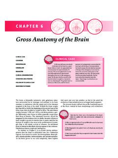
1 11 Human Anatomy and Physiology ILaboratoryGross Anatomy of the Brain and Cranial NervesThis lab involves the exercise entitled gross Anatomy of the Brain and Cranial Nerves . Complete the Review Sheet for the exercise and take the related quiz. There is also a video of the dissection of a sheep on the sound icon for the audio file (mp3 format) for each slide. There is also a link to a dowloadable mp4 video which can be played on an sulcusConvolutions: Sulcus or fissure = a groove;Gyrus = a raised : Sulcus or fissure = a groove;Gyrus = a raised sulcusFrontal lobeParietal lobeTemporal lobeOccipital lobeParieto-occipital fissureThe Cerebral CortexPre-central gyrusPost-central gyrus33 Lateral View of BrainCerebellum Frontal lobeTemporal lobeOccipital lobeParietal lobeLateral sulcus44 Cerebrum, Superior ViewCentral sulcusPrecentral gyrusPostcentral gyrusLongitudinal fissurePia mater follows all convolutions, contains blood GranulationsFalxcerebriThe white arachnoidgranulations are where cerebrospinal fluid is reabsorbed.
2 66 Olfactory bulbOptic nerve optic chiasma optic tractOculomotor n. Trigeminal n. Abducens n. Vestibulocochlear Accessory View of Brain w/ Cranial Nerves60 The Cranial NervesOn Old Olympus Towering Top AFinn And German Viewed Some OlfactoryII OpticIII OculomotorIV TrochlearV TrigeminalVI AbducensVII FacialVIII AcousticIX GlossopharyngealX VagusXI Spinal AccessoryXII vestibulocochlearand statoacousticThis rhyme gives you the first letters of the twelve cranial nerves in order. There are other rhymes that work, take your pick. You must learn the names, numbers (always use Roman numerals), and functions. There is no need to learn a rhyme for whether they are motor or sensory. Knowing their functions will tell you if they are motor or sensory. And the fact is that, while some are sensory only, all of the motor nerves have sensory proprioceptivefibers, despite the rhyme and the table in Marieb.
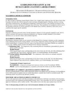
3 77 Ventral Brain, Posterior ViewHypoglossal (12th) Nerve Spinal Accessory (11th) Nerve Pons Medulla Abducens n. 88 Ventral view of Brain with Cranial NervesOlfactory bulbOptic chiasmaOculomotor , Ventral View1)Inferior Frontal Lobe, 2)Temporal Lobe, 3)Pons, 4)Medulla Oblongata, 5)Left CerebellarHemisphere, 6)Right CerebellarHemisphere1234561010 Sagittal Section of BrainCorpus callosumFornix Septum pellucidumIntermediate mass of thalamusHypothalamus Pons Medulla Choroid plexusPineal bodyCorpora quadrigeminaThird ventricleCerebellum Arbor vitaeCerebral aqueduct 4thventricle1111 Sagittal Section of Human Brain1) Cerebellum, 2) Pons, 3) Medulla, 4) Midbrain, 5) Mammillarybody, 6) Optic chiasma, 7) corpus callosum, 8) Septum pellucidum, 9) Cingulate gyrus, 10) Fornix, 11) Thalamus, 12)
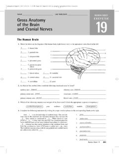
4 Section of Midbrain and DiencephalonCorpus callosumSeptum pellucidumChoroid plexusFornix Corpora quadrigeminaThalamus Hypothalamus Cerebral aqueduct4thventriclePons IM1313 Sheep Brain Dissection: Removal of the Dura Mater1414 Dura Mater from the Sheep Brain1515 Sheep Brain Dorsal View1616 Sheep Brain Cerebellum1717 Sheep Brain: The Corpora Quadrigemia1818 Sheep Brain Ventral View1919 Sheep Brain Sagittal Section2020 Cerebellar Purkinje CellsInterneuron cell bodiesDendrites Axon to cerebellaroutflowPurkinje cells are the largest and most distinguishing cells of the cerebellum. They have numerous dendrites and an axon which is the beginning of cerebellar s TangleThis is a neurofibrillary "tangle" of Alzheimer's disease. The tangle appears as long pink filaments in the cytoplasm. They are composed of cytoskeletalintermediate s Tangle, Silver StainThe characteristic microscopic findings of Alzheimer's disease include "senile plaques" which are collections of degenerative presynaptic endings along with astrocytes and microglia.
5 These plaques are best seen with a silver stain, as seen here in a case with many plaques of varying Protocol for Spinal Nerves and Reflexes1) Complete the Review Sheets for the exercise on the gross Anatomyof the Brain and Cranial Nerves2) Take the related quiz for the Brain and Cranial ) View the cadaver video showing dissection of the sheep brain.