Transcription of Human Anatomy and Physiology II Laboratory - …
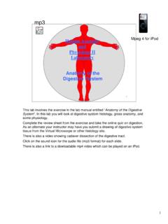
1 11 Human Anatomy and Physiology IILaboratory The Respiratory SystemThis lab involves two exercises in the lab manual entitled Anatomy of the Respiratory System and "Respiratory System Physiology ".In this lab you will look at lung histology, gross Anatomy , and Physiology . Complete the review sheets from the exercise and take the online quiz on respiration, As an alternate your instructor may have you submit a drawing of lung tissue from the Virtual Microsocpe or other histology site. Use the PhysioEX software to measure an analyze respiratory volumes.
2 There is also a video showing cadaver dissection of the respiratory on the sound icon for the audio file (mp3 format) for each slide. There is also a link to a dowloadable mp4 video which can be played on an TractUpper respiratory division Respiratory tree (Lower respiratory division)Nasal cavityTrachea 1oBronchi Pleurae Diaphragm Ribcage (thoracic wall)Larynx Pharynx The respiratory tract can be thought of as consisting of two parts: the upper respiratory tracthas the nasal cavity, and the pharynx; The lower respiratory tracthas the larynxand the respiratory treefrom the tracheathrough the various divisions of bronchito the bronchioles.
3 Essentially the upper division is where the cilia beat down to move mucus down to the throat to be swallowed, and the lower division is where cilia be up to move mucus to the throat to be Respiratory TractSee the Lab Manual for items you are responsible for inthe Upper Respiratory LarynxTracheaAnterior viewThyroid cartilageHyoid boneCricoid cartilageepiglottisMispronunciation of the larynxis an anatomical pet peeve. It is pronounced lair-inks, and consists of anumber of cartilages along with the ligaments which connect them.
4 The larynx is connected to the hyoid bone by the thryrohyoid ligament. The thryroid cartilageis the largest and its anterior prominence is the Adam s Apple .55 Sagittal Section of the LarynxTracheaPomum Adamii(Adam s Apple)Hyoid boneCricoid cartilageEpiglottis ArytenoidcartilageVocal folds (true vocal cords)Vestibular folds (false vocal cords)Arytenoid musclesThyroid cartilageWhen you swallow the hyoid bone lifts up and this causes the cartilaginous epiglottisto hinge backwards, guarding the opening into the glottis to prevent aspiration.
5 The glottisis the opening between the vocal folds, which are the vocal cords. The vocal folds are guarded by the vestibular folds. The arytenoid cartilage,controlled by arytenoid muscles, swivels to regulate tension on the vocal folds in producing the pitch of the voice. 66 Respiratory Tree(Dissected Specimen)Thyroid glandPrimary bronchiThyroid cartilageCricoidcartilageA dissected cadaver specimen of the larynx with its attachment to the respiratory , anterior viewThyroid cartilageCricothyroid muscleCricoid cartilageThe many small muscles found attached to the larynx have been removed from this specimen, along with the thyroid.
6 Posterior viewEpiglottis hyoid boneSuperior horn of thyroid cartilageArytenoidmusclesCricoid cartilageAryepiglotticfold and muscleCricoarytenoid musclePiriform recessMembraneouspart of tracheaThe epiglottis is not a separate leaf-like structure as it is often pictured, but rather is attached to the aryepiglottic fold to form more of a trumpet , sagittal sectionEpiglottis ArytenoidmuscleCricoidcartilageTrachea Cricoid cartilageVocalis muscleVocal foldventricleVestibular foldThyroid cartilageCadaver larynx in sagittal , superior viewepiglottisVestibular foldVocal foldAryepiglottic foldInterarytenoidnotchGlottis A view of the glottis from above.
7 When sound is produced the vocal folds tighten and loosen to produce different pitches, controlled by the arytenoidcartilages and arytenoid Cartilages, anterior viewEpiglottis Lesser cornuGreater cornuBody Hyoid boneThyroid cartilageThyroepiglottic ligamentCornus elasticusCricothyroid ligamentCornus elasticusCricoid cartilageWhen the soft tissues are removed, the cartilages of the larynx are Cartilages, posterior viewThyrohyoid ligamentLesser cornu of hyoid boneGreater cornu of hyoid boneSup. Cornu of thyroid cartilageCorniculate cartilageArytenoid cartilagePost.
8 JointEpiglottis Trachea Thyroepiglottic cartilageCricoarytenoid jointCricoid Trachea & EsophagusEsophagus Membraneousportion of trachea (trachealis muscle)C-ring glandsFibrous coveringA cross section of the trachea at the level of one of the C-ring cartilages shows how the esophagus fits into the membraneous portion of the trachea. This permits swallowing of a bolus of food. The trachealis muscle actually is a complete layer around the trachea, but is thicker at the posterior portion. Within the submucosa of the trachea lie seromucous glands.
9 The mucous they secrete helps to remove particulates through the action of the ciliated lining tissue (PCCE).1414 The Respiratory TreeCricoid cartilageTracheal cartilages2 Primary bronchi (to each lung)Subsegmental bronchi large and small, about 20 Secondary bronchi (to each lobe)18 Tertiary bronchi (to bronchopulmonarysegments)This is the bronchial portion of the respiratory zone: mucosa lined, allows no gas exchange with zone: thin walled simple squamous epithelium, allows gas exchange with ZoneThe large bronchiolesand terminal bronchiolesare still part of the conducting zone, which gets the air to and from the internal alveolar sacsof the lungs.
10 The respiratory bronchiolesand alveolar sac systemsare the respiratory zone, the part which allows gas transport by diffusion between the lungs and the into Respiratory BronchioleThe respiratory zone structures are composed of simple squamousepitheliumfor BronchusHyaline cartilage platePseudostratified ciliated columnar epitheliumSmooth muscleAlso see photos available on The Virtual MicroscopeA bronchus, at whatever level, is lined with pseudostratified ciliated columnar epithelium. Smooth muscle and a small amount of cartilage are also Smooth muscleAlveolus Capillaries Elastic fibersAlveolar sac systems(the sacs and their alveoli and ducts which lead to them) are simple squamous epithelium.