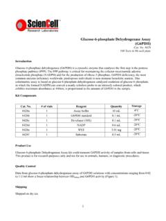
Transcription of Human Periodontal Ligament Fibroblasts
1 Human Periodontal Ligament Fibroblasts (HPLF) Catalog #2630 Cell Specification The Periodontal Ligament is connective tissue located between the cementum of the teeth and the alveolar bone of the mandibula. It plays an integral role in the maintenance and regeneration of Periodontal tissue. The cells responsible for maintaining this tissue are thought to be Fibroblasts , which can be either multipotent or composed of heterogeneous cell populations that differ in their function. Despite the similar morphology with gingival Fibroblasts , Periodontal Ligament Fibroblasts (PLF) appear to display distinct functional activities in the maintenance of tissue integrity [1]. It is known that PLF produce osteoblast-related extracellular matrix proteins and show higher alkaline phosphatase activity compared with gingival Fibroblasts [2].
2 PLF actively participate in immune and inflammatory events in Periodontal diseases [3]. HPLF from ScienCell Research Laboratories are isolated from Human Periodontal tissue. HPLF are cryopreserved at passage one and delivered frozen. Each vial contains >5 x 10⁵ cells in 1 ml volume. HPLF are characterized by their spindle morphology and immunofluorescence with antibodies specific to fibronectin. HPLF are negative for HIV-1, HBV, HCV, mycoplasma, bacteria, yeast and fungi. HPLF are guaranteed to further expand for 15 population doublings under the conditions provided by ScienCell Research Laboratories. Recommended Medium It is recommended to use fibroblast Medium (FM, Cat. #2301) for the culturing of HPLF in vitro.
3 Product Use HPLF are for research use only. They are not approved for Human or animal use, or for application in in vitro diagnostic procedures. Storage Upon receiving, directly and immediately transfer the cells from dry ice to liquid nitrogen and keep the cells in liquid nitrogen until they are needed for experiments. Shipping Dry ice. References [1] Han X, Amar S. (2002) Identification of genes differentially expressed in cultured Human Periodontal Ligament Fibroblasts vs. Human gingival Fibroblasts by DNA microarray analysis. J Dent Res. 81(6):399-405. [2] Murakami Y, Kojima T, Nagasawa T, Kobayashi H, Ishikawa I. (2003) Novel isolation of alkaline phosphatase-positive subpopulation from Periodontal Ligament Fibroblasts .
4 J Periodontol. 74(6):780-6. [3] Takashiba S, Naruishi K, Murayama Y. (2003) Perspective of cytokine regulation for Periodontal treatment: fibroblast biology. J Periodontol. 74(1):103-10. Instructions for culturing cells Caution: Cryopreserved cells are very delicate. Thaw the vial in a 37oC water bath and return the cells to culture as quickly as possible with minimal handling! Initiating the culture: 1. Prepare a poly-L-lysine-coated culture vessel (2 g/cm2, T-75 flask is recommended). Add 10 ml of sterile water to a T-75 flask and then add 15 l of poly-L-lysine stock solution (10 mg/ml, Cat. #0413). Leave the vessel in a 37oC incubator overnight (or for a minimum of one hour). 2. Prepare complete medium.
5 Decontaminate the external surfaces of medium bottle and medium supplement tubes with 70% ethanol and transfer them to a sterile field. Aseptically transfer supplement to the basal medium with a pipette. Rinse the supplement tube with medium to recover the entire volume. 3. Rinse the poly-L-lysine-coated vessel twice with sterile water and then add 15 ml of complete medium. Leave the vessel in the sterile field and proceed to thaw the cryopreserved cells. 4. Place the frozen vial in a 37oC water bath. Hold and rotate the vial gently until the contents completely thaw. Promptly remove the vial from the water bath, wipe it down with 70% ethanol, and transfer it to the sterile field.
6 5. Carefully remove the cap without touching the interior threads. Gently resuspend and dispense the contents of the vial into the equilibrated, poly-L-lysine-coated culture vessel. A seeding density of 5,000 cells/cm2 is recommended. Note: Dilution and centrifugation of cells after thawing are not recommended since these actions are more harmful to the cells than the effect of residual DMSO in the culture. It is also important that cells are plated in poly-L-lysine-coated culture vessels to promote cell attachment. 6. Replace the cap or lid of the culture vessel and gently rock the vessel to distribute the cells evenly. Loosen cap, if necessary, to allow gas exchange. 7. Return the culture vessel to the incubator.
7 8. For best results, do not disturb the culture for at least 16 hours after the culture has been initiated. Refresh culture medium the next day to remove residual DMSO and unattached cells, then every other day thereafter. Maintaining the culture: 1. Refresh supplemented culture medium the next morning after establishing a culture from cryopreserved cells. 2. Change the medium every three days thereafter, until the culture is approximately 70% confluent. 3. Once the culture reaches 70% confluency, change medium every other day until the culture is approximately 90% confluent. Subculturing: 1. Subculture when the culture reaches 90% confluency or above. 2. Prepare poly-L-lysine-coated culture vessels (2 g/cm2) one day before subculture.
8 3. Warm complete medium, trypsin/EDTA solution (T/E, Cat. #0103), T/E neutralization solution (TNS, Cat. #0113), and DPBS (Ca++- and Mg++ -free, Cat. #0303) to room temperature. We do not recommend warming reagents and medium in a 37oC water bath prior to use. 4. Rinse the cells with DPBS. 5. Add 8 ml of DPBS and then 2 ml of T/E solution into flask (in the case of a T-75 flask). Gently rock the flask to ensure complete coverage of cells by T/E solution. Incubate the flask in a 37oC incubator for 1 to 2 minutes or until cells completely round up. Use a microscope to monitor the change in cell morphology. 6. During incubation, prepare a 50 ml conical centrifuge tube with 5 ml of fetal bovine serum (FBS, Cat.)
9 #0500). 7. Transfer T/E solution from the flask to the 50 ml centrifuge tube (a small percent of cells may detach) and continue to incubate the flask at 37oC for another 1 to 2 minutes (no solution in the flask at this moment). 8. At the end of incubation, gently tap the side of the flask to dislodge cells from the surface. Check under a microscope to make sure that all cells detach. 9. Add 5 ml of TNS solution to the flask and transfer detached cells to the 50 ml centrifuge tube. Rinse the flask with another 5 ml of TNS to collect the residual cells. 10. Examine the flask under a microscope for a successful cell harvest by looking at the number of cells being left behind; there should be less than 5%.
10 Note: Use ScienCell T/E solution that is optimized to minimize cell damages due to over trypsinization. 11. Centrifuge the 50 ml centrifuge tube at 1000 rpm for 5 minutes. Resuspend cells in culture medium. 12. Count and plate cells in a new poly-L-lysine-coated culture vessel with the recommended cell density. Caution: Handling Human derived products is potentially biohazardous. Although each cell strain tests negative for HIV, HBV and HCV DNA, diagnostic tests are not necessarily 100% accurate, therefore, proper precautions must be taken to avoid inadvertent exposure. Always wear gloves and safety glasses when working with these materials. Never mouth pipette. We recommend following the universal procedures for handling products of Human origin as the minimum precaution against contamination [1].