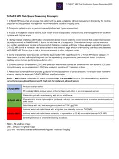
Transcription of III. REPORTING SYSTEM - ACR
1 ACR BI-RADS ATLAS BREAST MRIA merican College of Radiology 125 MAGNETIC RESONANCE IMAGINGIII. REPORTING SYSTEM2013126 American College of Radiology MAGNETIC RESONANCE IMAGINGACR BI-RADS ATLAS BREAST MRIA merican College of Radiology 127 MAGNETIC RESONANCE IMAGINGA. REPORT ORGANIZATIONThe REPORTING SYSTEM should be concise and organized.
2 Any pertinent clinical history that may af-fect scan interpretation, as well as any MRI acquisition techniques (including postprocessing informa-tion) that may affect the scan interpretation, should be described. The breast MRI report should first describe the amount of fibroglandular tissue and the background parenchymal enhancement (BPE). Abnormal enhancement (unique and separate from the BPE) is described based on morphology, distribution, and kinetics. Results from any physiologic or parametric imaging should be described. An assessment is rendered that includes the degree of concern and any recommendation(s).
3 Benign findings need not be reported, especially if the interpreting physician is concerned that the referring clinician or patient might infer anything other than absolute confidence in benignity. Table 2. Report OrganizationReport Structure1. Indication for examination2. MRI technique 3. Succinct description of overall breast composition4. Clear description of any important findings5. Comparison to previous examination(s)6. Assessment7. Management1. INDICATION FOR EXAMINATION Provide a brief description of the indication for examination. For example, this may be high-risk screening, follow-up of a probably benign lesion, follow-up of cancer treated with neoadjuvant chemotherapy, or evaluation of the newly diagnosed cancer patient.
4 As background parenchymal enhancement can be affected by cyclical hormonal changes, it may be helpful to include menstrual history. If the patient is pre- menopausal , the week of the menstrual cycle may be important information to aid in interpretation. Current therapy (neoadjuvant, adju-vant, hormonal, or radiation therapy ) for breast cancer treatment in the pre-or postsurgical setting may be important information and may inform exam interpretation. The indication for examination should contain a concise description of the patient s clinical his-tory, including:a. Reason for performing the exam ( , screening, staging, problem solving)b.
5 Clinical abnormalities, including size, location, and duration i. Palpable finding ii. Nipple discharge iii. Other pertinent clinical findings or history2013128 American College of Radiology MAGNETIC RESONANCE IMAGINGc. Previous biopsies i. Biopsy type ii. Biopsy location iii. Benign or malignant pathology (cytology or histology)d. Hormonal status if applicable i. Pre- or post- menopausal ii. Menstrual cycle phase (second week or other) or last menstrual period iii. Peripartumiv. Exogenous hormone therapy , tamoxifen, aromatase inhibitors, or other hormones or medications/herbs/vitamins that might influence MRI2.
6 MRI TECHNIQUE Give a detailed description of the technical factors of how the MRI examination was obtained. At a minimum, a bright-fluid sequence of both breasts should be obtained. Pre- and post-gadolinium T1-weighted images should be obtained, preferably with fat suppression, simultaneously of both breasts. Subtraction imaging may be desired as well as other as processing techniques and para-metric analysis. Elements of this description routinely include: a. Right, left, or both breasts b. Location of markers and their significance (scar, nipple, palpable lesion, etc.) c. Weighting i.
7 T1 weighted ii. T2 weighted iii. Fat saturation iv. Scan orientation and plane v. Other pertinent pulse sequence features d. Contrast dose i. Name of contrast agent ii. Dosage (mmol/kg) and volume (in cc) iii. Injection type: bolus or infusion iv. Timing (relationship of bolus injection to scan start time and scan length)v. If multiple scans: number of postcontrast scans and acquisition techniques of each (how fast, how many slices, and slice thickness) e. Postprocessing techniques as applicable i. MPR/MIP ii. Time/signal intensity curves iii. Subtraction iv.
8 Other techniquesACR BI-RADS ATLAS BREAST MRIA merican College of Radiology 129 MAGNETIC RESONANCE IMAGING 3. SUCCINCT DESCRIPTION OF OVERALL BREAST COMPOSITION This should include an overall description of the breast composition, including: a. The amount of FGT that is presentTable 3. Breast Tissue Fibroglandular Tissue (FGT)Amount of Fibroglandular Tissuea. Almost entirely fatb. Scattered fibroglandular tissue c. Heterogeneous fibroglandular tissued.
9 Extreme fibroglandular tissueThe four categories of breast composition (Table 3) are defined by the visually estimated content of FGT within the breasts. If the breasts are not of apparently equal amounts of FGT the breast with the most FGT should be used to categorize breast composition. Although there may be considerable variation in visually estimating breast composition, categorizing based on percentages (and specifically into quartiles) is not recommended. We recognize that quantification of breast FGT volume on MRI may be feasible in the future, but we await publication of robust data before endorsing percentage recommendations .
10 We urge the use of BI-RADS terminology instead of numbers to classify breast FGT in order to eliminate any possible confusion with the BI-RADS assessment categories, which are 262 Almost entirely 263 Scattered fibroglandular American College of Radiology MAGNETIC RESONANCE IMAGINGF igure 264 Heterogeneous fibroglandular 265 Extreme fibroglandular The amount of background parenchymal enhancement in the imageTable 4. Breast Tissue Background Parenchymal Enhancement (BPE)Background Parenchymal Enhancementa. Minimalb. Mildc. Moderated. MarkedThe four categories of BPE (Table 4) are defined by the visually estimated enhancement of the FGT of the breast(s).


