Transcription of Immunocytochemistry (ICC) - Novus Biologicals
1 Learn more | more | (ICC) HandbookConjugateExcitation/EmissionEmis sionColorLaser (Excitation Source)NotesDyLightTM 405400/420 VioletViolet (405 nm)Bright and photostableAlexa Fluor 405401/421 VioletViolet (405 nm)Best when used with more abundant targetsDyLightTM 350353/432 Violet-BlueUltraviolet (355 nm)Can be used with DyLightTM 488, 594 and 647 in multiplexingAlexa Fluor 350346/442 Violet-BlueUltraviolet (355 nm)Often used with Alexa Fluor 488, 594 and 647 in multiplexing, best for high-abundance targetsDyLightTM 488493/518 GreenBlue (488) Brighter, photostable replacement for FITC; not suitable for use with GFPA lexa Fluor 488495/519 GreenBlue (488) Photostable over a broad pH range; replaces FITCFITC495/519 GreenBlue (488) Small organic fluorophore; cannot be used with DyLightTM 488, Alexa Fluor 488 or GFPDyLightTM 405LS397/572 YellowViolet (405 nm)Superior alternative to Pacific Orange; good choice for multicolor applications on the violet laserAlexa Fluor 546556/573 YellowYellow-Green (561 nm)Photostable over a broad pH range; brighter than Cy3 DyLightTM 550562/576 YellowYellow-Green (561 nm)PE565/578 YellowYellow-Green (561 nm)Subject to photobleaching.
2 Can be excited by the 488, 532, and 561nm lasers on flow cytometersTexas Red 595/613 OrangeYellow-Green (561 nm)Very bright fluorescence; use a tunable dye laserto avoid leaking when multiplexed with PEAlexa Fluor 594590/617 OrangeYellow-Green (561 nm)Better photostability than Texas RedDyLightTM 650654/673 RedRed (633 nm)APC650/660 RedRed (633 nm)Bright fluorescent protein; do not use with DyLightTM 650 due to overlapping emission spec-traAlexa Fluor 647650/665 RedRed (633 nm)Extremely photostable, good replacement for Cy5 or APCCy5TM647/665 RedRed (633 nm)Some fluorescence quenching when conjugatedDyLightTM is a registered trademark of Thermo Fisher Scientific Inc.
3 Alexa Fluor and Texas Red are registered trademark of Life Science Technologies Corpora-tion. Cy5TM is a trademark of GE 21-222324 TABLE OF CONTENTSI ntroduction Basic ICC/IF Protocol Multicolor ICC/IF Why use IF based detection Multicolor ICC/IF - limitations Considerations Before You Start Autofluorescence Fluorochrome selection Sample Preparation How important is it Coverslip preparation and coating Fixation Why fix the cells Types of fixatives Permeabilization Blocking The significance of blocking Blocking buffers Antibodies Reactivity Specificity Clonality Host Application compatibility Concentration and incubation time Antibody Washes Detection
4 Methods Direct and indirect detection Which method of detection is better Controls Counterstaining and Mounting Counterstaining Mounting Multicolor ICC/IF Protocol (bench-top) General steps: cell culture, fixation, permeabilization, and blocking Sequential incubations with unlabeled antibodies Simultaneous incubation with directly labeled primary antibodies Simultaneous incubation with unlabeled primary antibodies Pros and cons of multi-staining methods Troubleshooting Tips Common Organelle Markers for ICC/IF Reagent Recipes Conjugated Antibodies Conjugated antibodies Antibody labeling kitsLearn more | 1 Immunocytochemistry (ICC) refers to immunostaining of cultured cell lines or primary cells including smears, swabs, and aspirates.
5 ICC offers a semi-quantitative means of analyzing the relative abundance, conformation, and subcellular localization of target antigens. Traditional ICC techniques use chromogenic detection in which enzyme conjugated antibodies convert chromogen substrates to a colored precipitate at the reaction site. However chromogenic detection has lost favor with the advent of immunofluorescent labels. In Immunocytochemistry /immunofluorescence (ICC/IF) assays, the cellular antigens are visualized using either fluorochrome-conjugated primary antibodies (direct detection) or a two-step method (indirect detection) involving an unlabeled primary antibody followed by a fluorochrome-conjugated secondary antibody.
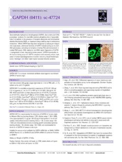
6 By combining different fluorochrome-labeled antibodies, multiplex ICC/IF can detect several antigens in the same ICC is often used interchangeably with ICC/IF ( Immunocytochemistry / immunofluorescence) and another related term, IF, significant differences exist between them in reference to the method of detection and starting sample types involved. Some of the key differences between ICC, ICC/IF and IF are illustrated in the table and images below: Assay TerminologySampleDetectionICC (traditional)CellsEnzyme-labeled antibodies and corresponding chromogen substrates (chromogenic detection)ICC/IFCellsFluorochrome-labele d antibodies (immunofluorescent detection)IFTissues or CellsImmunofluorescent detection, can be applicable to ICC and/or IHC Figure 1.
7 Examples of staining using traditional ICC, ICC/IF, and IHC with IF based detectionIRAK4 was detected in K562 cells using 10 g/ml of IRAK4 antibody [NBP1-77231] and HRP conjugated secondary antibody. Cells were stained with DAB (brown) and nuclei were counterstained with hematoxylin (blue). IRAK4 was detected in THP 1 cells using 15 g/ml IRAK4 antibody [AF3919]. The signal was de-veloped using NorthernLights 557-conjugated secondary antibody (red) and nuclei were counterstained with DAPI (blue). GAP43 was detected in a formalin fixed paraffin embedded section of mouse embryo using 1ug/ml of GAP-43 antibody [NB300-143] followed by Alexa Fluor 488 labeled secondary.
8 DAPI was used as a nuclear counterstain. Traditional ICCICC/IFExample of IF in IHCL earn more | more | ICC/IF Protocol A standard ICC/IF protocol involves fixation, permeabilization, blocking, immunolabeling, counterstaining, and microscopic imaging of stained cells (see the flow chart in Figure 2). Each step of the ICC/IF protocol requires optimization as experimental variables in each step can significantly impact staining outcome. Chemical crosslinkers(formaldehyde)FixationPermea bilization(Triton x100)Blocking(normal serum)Antibody incubationDirect IF detectionIndirect IF detectionPrimary antibody(labeled)Primary antibody(unlabeled)Precipitants(methanol , acetone)Secondary antibody(labeled)Counterstaining(DAPI, Phalloidin, etc.)
9 MountingMicroscopic analysisCells(cultured or smears)Sample PreparationImmunostainingAnalysisFigure 2. General protocol steps in an ICC/IF assayLearn more | ICC/IFWhy use IF based detection? The benefit of fluorescence detection is the ability to simultaneously detect multiple structural and functional molecules (multiplex analysis) with high contrast and visibility. Due to the narrow spectral properties of most fluorochromes, the expression of multiple proteins can be detected at the same time and better distinguished relative to chromogen based detection methods, which demonstrate broad spectral properties.
10 Often fluorescent imaging is used to compare the subcellular localization or distribution of two or more fluorescently labeled molecules. These comparisons can then be used to understand protein function, including intracellular transport, response to biological stimuli or disease, and target protein co-localization with a marker or organelle. Unlike fluorescence detection, the broad emission spectra of chromogens make it difficult to distinguish individual colors that coincide on the same structure/organelle (see Figure 3). Thus, the relative simplicity of multiplex analysis and ability to clearly detect co-localized antigens make fluorescence detection more desirable than chromogen based detection methods in ICC/IF - limitations?