Transcription of Immunohistochemistry (IHC) - Novus Biologicals
1 Learn more | more | (IHC) Handbook Variables Influencing Experimental Design VariablesFactors*TissueSpecies, Type, Section Size and ThicknessTargetAbundance and Subcellular LocalizationEpitopeConformation Dependent Availability and Post-translational ModificationsSample PreparationParaffin-embedded or FrozenFixation MethodPerfusion or ImmersionFixativeAldehydes (Crosslinking), Alcohols (Precipitating), or AcetoneBlocking BufferNormal Serum, BSA, Casein, Various Detergents, and Salt ConcentrationsAntigen Retrieval MethodNone, Heat-induced Epitope Retrieval (HIER), and Protease-induced Epitope Retrieval (PIER)PermeabilizationTriton-X 100, Tween-20, or SaponinDetection MethodDirect, Indirect, or Signal Amplification MethodsPrimary AntibodyMonoclonal or PolyclonalSecondary AntibodySpecies Reactivity and LabelMultiplexSimultaneous or Sequential Antibody Addition, and Antibody SpeciesImaging MethodFluorescence or ChromogenicLabelFluorochromes (numerous options)Chromogenic Reagents: Enzymes (HRP, AP) and Substrates (DAB, AEC, NBT/BCIP)CounterstainFluorescence: DAPI, Hoechst 33342 Chromogenic.
2 Hematoxylin, Fast Green FCF* The above table is not intended to be exhaustive but rather summarizes common factors influencing IHC experimental design at to IHCS ample Preparation and Fixation Key Differences of Sample Preparation Standard Fixatives What is the Difference Between Paraformaldehyde, Formaldehyde, and Formalin? Sample Preparation and Sectioning ProtocolsEpitope (Antigen) Retrieval Is Antigen Retrieval Necessary? HIER Protocol PIER ProtocolBlocking Non-Specific Binding Endogenous Activity and Reactive EpitopesAntibodies and Detection Methods Key Differences of Antibody Clonality Detection Methods Signal Amplification Methods Which Detection Method is Best for Multiplex IHC?
3 IHC Controls Positive Tissue Control Negative Tissue Control Tissue Artifact Control Antibody ControlsComplementary Techniques Western Blotting RNA in situ HybridizationIHC Staining Protocol Deparaffinization and Rehydration Protocol Permeabilization IF Staining Protocol Chromogenic Staining ProtocolIHC WorkflowTroubleshooting Guide No or Poor Signal High Background Poor Tissue Morphology Uneven or Non-specific StainingReference BuffersBio-Techne Support Products for IHC 12-56-78-910-1112-131415-1718-1920-22232 4 TABLE OF CONTENTSL earn more | 1 Immunohistochemistry (IHC) uses antibodies to detect cell and tissue proteins and provide semi-quantitative data about target protein expression, distribution, and localization.
4 Tissues are sectioned from fixed embedded ( IHC- paraffin or plastic) or frozen blocks (e. g. IHC-Frozen), and the sections are then probed with primary antibodies against the antigens of interest. Target expression can be evaluated with the corresponding labeled primary antibody (direct detection) or, more commonly, with the addition of labeled secondary antibodies (indirect detection). The label, either fluorescent or enzymatic, is used to visualize the antigen-antibody this multi-step application, staining may be influenced by multiple variables and requires the careful optimization of new assays for each target antigen. For example, when investigating a high abundance protein in formaldehyde-fixed tissue, the IHC protocol may include a heat-induced antigen retrieval step and use a directly labeled primary antibody.
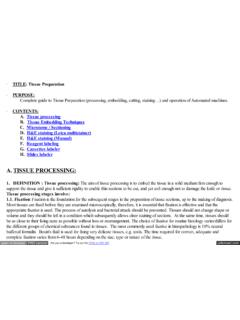
5 In another example, the optimal protocol for staining a low abundance protein in a methanol fixed, frozen liver section may require blocking of endogenous biotin and a signal amplification a number of variables can introduce artifacts or interfere with a successful outcome, appropriate controls are necessary for accurate interpretation of IHC results. This brief guide is intended to serve as a reference for researchers to understand, perform, and troubleshoot various IHC protocols during the development and optimization of new IHC of interest is stained and imaged with microscopeNuclear staining of islet cellsSubstrateSignalSecondary AntibodyPrimary AntibodyEnzymeAntigenSTAININGVISUALIZATI ONB asics of an IHC ExperimentLearn more | more | can be broadly classified into two forms based on the type of tissue processing involved: IHC- formalin-fixed, paraffin -embedded (FFPE) and IHC-frozen (Fr).
6 Often the preservation method is closely associated with the type of fixation. Formalin-fixed tissues are commonly paraffin -embedded following fixation, while frozen tissue sections can be fixed with formaldehyde or alcohol prior to or following cryosectioning. Sample Preparation and Fixation IHC-FFPEIHC-FrFixationPerformed before embedding into paraffin be performed before or after FixativeFormaldehydeFormaldehyde or AlcoholsSectioningMicrotome, 4-10 m sectionsCryostat/Cryotome, 5-20 m sectionsStorageIdeally fresh sections should be cut after 4 weeks due to loss of antigenic cut sections should not be used after 1 month. For long term storage (several years), coating of slide in paraffin is recommended.
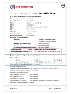
7 Short-term-1 year at -80 AdvantageEase of handling and preserves structural morphology. Blocks can be stored long enzyme & antigen function. Useful for study of post-translationally modified protein, DNA, or RNA. Major LimitationsVariable fixation times. Fixation can mask optimal for studying structural morphology. Ice crystals can impact tissue Differences of Sample PreparationFixation preserves the histologically relevant morphology of tissues and antigenicity of target molecules. For the best results, tissue should undergo rapid and uniform fixation. Whole animal perfusion (Figure 1A), using an animal s vascular network to disperse the fixative solution, is the preferred method for tissues from small animals including mice and rats.
8 Alternatively, dissected tissue can be directly immersed in the fixative immediately after tissue collection (Figure 1B). For fixation byimmersion, tissue should be cut into 10 mm thick pieces and then immersed in fixative solution, 50-100X the tissue volume, for effective diffusion of the fixative. ABFigure 1. Fixation Methods: Perfusion (A) vs. Immersion (B) Learn more | Preparation and Fixation Standard Fixatives4% formaldehyde in phosphate-buffered saline (PBS) is the most common fixative for preserving protein targets in tissues. Formaldehyde reacts with amino groups in proteins to form methylene bridges that crosslink proteins in tissue sections. These molecular cross-links can mask protein epitopes from antibody binding and may require the addition of an antigen retrieval step for proper IHC staining.
9 Additionally, formaldehyde-mediated tissue fixation has been shown to induce translocation of phosphorylation-dependent epitopes from the membrane to the predominant alcohols used for fixation are 70% methanol and 80% ethanol. Alcohols work by removing and replacing water molecules in tissue, which can destabilize hydrophobic bonds and alter the tertiary structure of proteins. This also causes the precipitation of soluble proteins, making alcohol-mediated fixation more appropriate for detection of membrane bound proteins. Aldehydes AlcoholsNo single fixation condition works for all target molecules and tissues. Finding the optimal fixative and fixation time for each antigen is critical to preserve tissue morphology and antibody binding capacity.
10 Any issues arising from incomplete fixation such as autolysis cannot be reversed or fully rectified in later is the difference between paraformaldehyde, formaldehyde, and formalin? Paraformaldehyde (PFA) is the polymerized form of formaldehyde and is not itself a fixing agent. Formaldehyde can be prepared by dissolving PFA in PBS using heat and sodium hydroxide (NaOH). Formalin refers to a saturated formaldehyde solution and some commercial formalin solutions include methanol as a stabilizer to prevent formaldehyde polymerization. A 10% formalin solution is equivalent to a formaldehyde is also used as a strong dehydrant and precipitant, typically applied to sections of snap-frozen tissues. Acetone fixation is generally mild and may be followed by fixation with alcohols or more | more | Preparation and Sectioning 01 PROTOCOL: Formalin-fixed, paraffin -embedded (FFPE)02030405 Fix the tissue of interest by immersing it in 10% neutral buffered formalin (4% formaldehyde) for 4-24 hours at room temperature.