Transcription of Indications by Appropriate Use Ratings
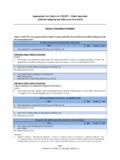
1 Appropriate Use Criteria for ICD/CRT Online Appendix Indications by Appropriate Use Ratings Table 1. Appropriate Indications (Median Score 7-9). Appropriate Indication Use Score (1-9). Section 1: Secondary Prevention CAD: VF or Hemodynamically Unstable VT Associated With Acute (<48 hours) MI. (Newly Diagnosed, No Prior Assessment of LVEF). Total Revascularization Completed After Cardiac Arrest VF or polymorphic VT during acute (<48 hours) MI. NSVT 4 days post MI. 3. A (7). Inducible VT/VF at EPS 4 days after revascularization LVEF 36-49%.
2 VF or polymorphic VT during acute (<48 hours) MI. NSVT 4 days post MI. 3. A (8). Inducible VT/VF at EPS 4 days after revascularization LVEF 35%. Obstructive CAD With Coronary Anatomy Not Amenable to Revascularization VF or polymorphic VT during acute (<48 hours) MI. 6. No EPS done A (7). LVEF 35%. CAD: VF or Hemodynamically Unstable VT <48 Hours (Acute) Post-Elective Revascularization No evidence for acute coronary occlusion, restenosis, preceding infarct, or other clearly 7. reversible cause A (7).
3 LVEF 35%. CAD: VF or Hemodynamically Unstable VT [No Recent MI ( 40 days) Prior to VF/VT and/or No Recent Revascularization ( 3 Months) Prior to VF/VT]. No identifiable transient and completely reversible causes 8. No need for revascularization identified by cath performed following VF/VT A (9). LVEF 50%. No identifiable transient and completely reversible causes 8. No need for revascularization identified by cath performed following VF/VT A (9). LVEF 36-49%. No identifiable transient and completely reversible causes 8.
4 No need for revascularization identified by cath performed following VF/VT A (9). LVEF 35%. No revascularization performed (significant CAD present at cath performed following VF/VT, 9. but coronary anatomy not amenable to revascularization) A (9). LVEF 50%. Page 1. American College of Cardiology Foundation No revascularization performed (significant CAD present at cath performed following VF/VT, 9. but coronary anatomy not amenable to revascularization) A (9). LVEF 36-49%. No revascularization performed (significant CAD present at cath performed following VF/VT, 9.)
5 But coronary anatomy not amenable to revascularization) A (9). LVEF 35%. Significant CAD identified at cath performed following VF/VT. 10. Complete revascularization performed after cardiac arrest A (7). LVEF 36-49%. Significant CAD identified at cath performed following VF/VT. 10. Complete revascularization performed after cardiac arrest A (7). LVEF 35%. Significant CAD identified at cath performed following VF/VT. 11. Incomplete revascularization performed after cardiac arrest A (7). LVEF 50%. Significant CAD identified at cath performed following VF/VT.
6 11. Incomplete revascularization performed after cardiac arrest A (8). LVEF 36-49%. Significant CAD identified at cath performed following VF/VT. 11. Incomplete revascularization performed after cardiac arrest A (9). LVEF 35%. CAD: VF or Hemodynamically Unstable VT During Exercise Testing Associated With Significant CAD. No revascularization performed (significant CAD present at cath performed following VF/VT, 12. but coronary anatomy not amenable to revascularization) A (9). LVEF 50%. No revascularization performed (significant CAD present at cath performed following VF/VT, 12.)
7 But coronary anatomy not amenable to revascularization) A (9). LVEF 36-49%. No revascularization performed (significant CAD present at cath performed following VF/VT, 12. but coronary anatomy not amenable to revascularization) A (9). LVEF 35%. Significant CAD identified at cath performed following VF/VT. 13. Complete revascularization performed after cardiac arrest A (7). LVEF 35%. Significant CAD identified at cath performed following VF/VT. 14. Incomplete revascularization performed after cardiac arrest A (7).
8 LVEF 50%. Significant CAD identified at cath performed following VF/VT. 14. Incomplete revascularization performed after cardiac arrest A (7). LVEF 36-49%. Significant CAD identified at cath performed following VF/VT. 14. Incomplete revascularization performed after cardiac arrest A (8). LVEF 35%. Page 2. American College of Cardiology Foundation No CAD: VF or Hemodynamically Unstable VT. Dilated nonischemic cardiomyopathy 15. A (9). LVEF 50%. Dilated nonischemic cardiomyopathy 15. A (9). LVEF 36-49%.
9 Dilated nonischemic cardiomyopathy 15. A (9). LVEF 35%. VF/Hemodynamically Unstable VT Associated With Other Structural Heart Disease 18. Myocardial sarcoidosis A (9). 20. Giant cell myocarditis A (8). Genetic Diseases with Sustained VT/VF. 22. Congenital Long QT A (9). 23. Short QT A (9). 24. Catecholaminergic Polymorphic VT A (9). 25. Brugada syndrome A (9). 26. ARVC with successful ablation of all inducible monomorphic VTs A (9). 27. ARVC with unsuccessful attempt to ablate an inducible VT A (9).
10 28. ARVC without attempted ablation A (9). 29. Hypertrophic cardiomyopathy A (9). No Structural Heart Disease (LVEF 50%) or Known Genetic Causes of Sustained VT/VF. Idiopathic VF With Normal Ventricular Function 32. No family history of sudden cardiac death A (9). 33. First degree relative with sudden cardiac death A (9). Syncope in Patients Without Structural Heart Disease Unexplained Syncope in a Patient With Long QT Syndrome 41. While on treatment with beta blockers A (9). 42. Not being treated with beta blockers A (7).
