Transcription of INTERNATIONAL CLINICAL DIABETIC …
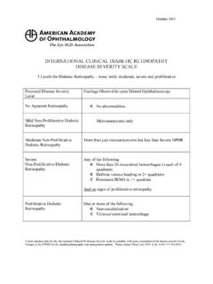
1 October 2002. INTERNATIONAL CLINICAL DIABETIC RETINOPATHY. DISEASE SEVERITY SCALE. DETAILED TABLE. Proposed Findings Observable upon Derivation from Risk Management Options*. Disease Dilated Ophthalmoscopy ETDRS Levels Assessment Severity Level No apparent No abnormalities Levels 10: DR Optimize medical therapy of Retinopathy absent glucose, blood pressure and lipids Mild Non- Microaneurysms Level 20: Very Optimize medical therapy of Proliferative only mild NPDR glucose, blood pressure and DIABETIC lipids Retinopathy Moderate More than just Levels 35,43: One year early Refer to an ophthalmologist Non- microaneurysms but moderate NPDR PDR: Optimize medical therapy of proliferative less than Severe less than 4:2.
2 1 glucose, blood pressure and DIABETIC NPDR One year high lipids Retinopathy risk Level 47: One year early Refer to an ophthalmologist moderate NPDR PDR Optimize medical therapy of less than 4:2:1 One year High glucose, blood pressure and Risk PDR: lipids Severe Non- Any of the following: 53A-E: severe One year risk Consider scatter (panretinal). Proliferative Extensive (>20) to very severe for early PDR: laser treatment for patients with DIABETIC intraretinal NPDR, 4:2:1 (severe type 2 diabetes Retinopathy hemorrhages in each of rule NPDR) Optimize medical therapy of 4 quadrants One year High glucose, blood pressure and Definite venous Risk PDR: lipids beading in 2+ (severe quadrants NPDR).
3 Prominent IRMA in (very 1+ quadrant severe NPDR). And no signs of proliferative retinopathy Proliferative One or more of the following: Levels 61, 65, Strongly consider scatter DIABETIC Neovascularization 71,75, 81,85: (panretinal) laser treatment , Retinopathy Vitreous/preretinal PDR, high-risk without delay for patients with hemorrhage PDR, very vitreous hemorrhage or severe or neovascularization within one advanced PDR disc diameter of the optic nerve head Optimize medical therapy of glucose, blood pressure and lipids * These management options are provided as general practice patterns of care. Individualized treatment plans will vary, based on several CLINICAL considerations and factors, based on the patient's circumstances, risk factors, systemic condition, etc.
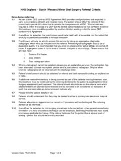
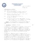
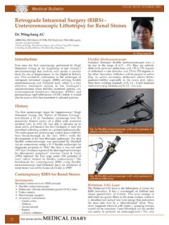
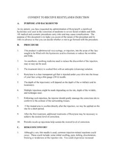
4 There are many modifiers or risk factors not included in this classification, but which are important in risk of disease progression and in managing individual patients. These factors should be taken into account by the clinician in decisionmaking, and in informing the patient and primary care physician/diabetologist. October 2002. PHOTO 2A: Hemorrhages of this severity in 4 quadrants = severe NPDR . October 2002. PHOTO 8A: IRMA of this severity in 1 quadrant = Severe NPDR . October 2002. Color DIABETIC Grading Standard Photographs Standard 2A-left Standard 2A-right Standard 8A-left Standard 8A-right Fundus Photograph Reading Center Reprinted with permission from the Fundus Photograph Reading Center Dept.
5 Of Ophthalmology & Visual Sciences University of Wisconsin - Madison Dept. of Ophthalmology & Visual Sciences University of Wisconsin - Madison October 2002. Detailed Discussion of treatment Recommendations (based on AAO DIABETIC Retinopathy Preferred Practice Pattern, 1998). Normal NPDR. The patient with a normal retinal exam should be re-examined annually because within 1 year, 5% to 10% of patients who are initially normal will develop DIABETIC retinopathy. Existing retinopathy will worsen by a similar percentage. laser surgery, color fundus photography, and fluorescein angiography are not indicated.
6 Mild NPDR. Moderate NPDR. Patients with retinal microaneurysms and hard exudates or other abnormalities should have a repeat examination within 6 to 12 months, because disease progression is common. In one study of Type 1 patients, 16% with mild retinopathy (hard exudates and microaneurysms only) progressed to proliferative stages within 4 years. laser surgery and fluorescein angiography are not indicated for this group of patients. Color fundus photography is not particularly helpful as a baseline for future comparison Severe NPDR. The categories of Severe NPDR, very severe NPDR and non high-risk PDR are combined for discussion because the ETDRS data showed their similar CLINICAL course, and subsequent recommendations for treatment are similar.
7 In eyes with severe NPDR, the risk of progression to proliferative disease is high. Half of the patients with severe NPDR will develop PDR within 1 year, and 15% will be high-risk PDR. For patients with very severe NPDR, the risk of developing PDR within 1 year is 75%, and 45% will be high-risk PDR. Therefore, these patients should be re-examined within 3 to 4 months. The value of laser surgery for this group of patients was studied by the ETDRS. The ETDRS compared early panretinal photocoagulation with deferral of photocoagulation, defined as careful follow-up (at 4-month intervals). and prompt panretinal photocoagulation if progression to high-risk PDR occurred.
8 Although the study did not provide definitive guidelines, the ETDRS suggested that panretinal photocoagulation is not recommended in eyes with mild or moderate NPDR, provided follow-up can be maintained. When retinopathy is more severe, panretinal photocoagulation should be considered and usually should not be delayed if the eye has reached the high-risk proliferative stage. Careful follow-up at 3 to 4 months is important: if the patient will not or cannot be followed closely, then laser photocoagulation may be indicated. If laser surgery is elected, full panretinal photocoagulation is a proven surgical technique that has been described in the literature.
9 laser photocoagulation may be indicated particularly when access to health care is difficult. Recent additional analyses of visual outcome in ETDRS patients with severe NPDR to non-high-risk PDR suggest that the recommendation to consider scatter photocoagulation prior to the development of high-risk PDR is particularly appropriate for patients with Type 2 diabetes. The risk of severe vision loss or vitrectomy was reduced by 50% in those who were treated early compared with the deferral until high-risk PDR developed. For patients with Type 1 diabetes, the timing of the scatter photocoagulation will depend on the compliance with follow-up, status and response to treatment of the fellow eye, impending cataract surgery, and/or pregnancy.
10 October 2002. The goal of laser treatment is to reduce the rate of visual loss. Preoperatively, the ophthalmologist should assess macular edema, discuss side effects of treatment and risks of visual loss, and obtain informed consent from the patient. If retrobulbar or peribulbar injections are used prior to photocoagulation, plans should be made to manage anticipated complications. Serious complications are rare, but they do occur. The ETDRS protocol provides detailed guidelines for treatment . Postoperatively, the eye should be patched if retrobulbar or peribulbar block was used. The patient should be seen at follow-up visits every 1 to 4 weeks until completion of panretinal photocoagulation, and then every 1 week to 4 months thereafter.