Transcription of Key Points - BAUS
1 Information about your procedure from The British Association of Urological Surgeons (BAUS) Published: June 2021 Leaflet No: 21/164 Page: 1 Due for review: June 2023 British Association of Urological Surgeons (BAUS) Limited This leaflet contains evidence-based information about your proposed urological procedure. We have consulted specialist surgeons during its preparation, so that it represents best practice in UK urology. You should use it in addition to any advice already given to you. To view the online version of this leaflet, type the text below into your web browser: Further general information about kidney stones can be found on the Patients section of the BAUS website under "I think I might have .. kidney stones". What does this procedure involve?
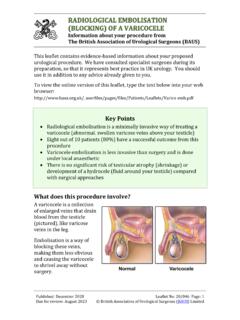
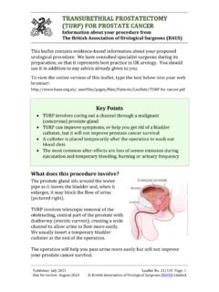
2 This involves puncturing your kidney through the skin of your back with a needle (using X-ray or ultrasound to guide the puncture) and stretching up a track into the kidney through which a telescope can be passed. We may need to puncture the kidney at more than one site to reach all your stone(s). Key Points The aim of this procedure is to fragment stones in the kidney telescopically using a keyhole approach to your kidney through a puncture in the skin of your back It is a major procedure and is usually reserved for larger stones or for patients with complex kidney anatomy We puncture the kidney with a needle, under ultrasound or X-ray guidance, and stretch up a track into the kidney through which we can pass a telescope The stone is broken up using a laser fibre, a lithoclast (small pneumatic drill) or an ultrasonic suction probe This procedure has largely eliminated the need for open surgery to remove kidney stones, because there is a similar stone clearance rate and recovery is much faster Published: June 2021 Leaflet No.
3 21/164 Page: 2 Due for review: June 2023 British Association of Urological Surgeons (BAUS) Limited We then fragment the stone(s) in your kidney using a laser, lithoclast (mechanical fragmenter) or ultrasound probe. We may leave a drainage tube in your kidney (nephrostomy) or an internal tubve (ureteric stent) at the end of the procedure and we usually put a catheter in your bladder. What are the alternatives? Observation stones smaller than 5mm in diameter can pass by themselves but sometimes get stuck within the kidney ; larger stones (greater than 7mm diameter) rarely pass Extracorporeal shockwave lithotripsy (ESWL) by firing shockwaves, generated under water, through your skin to break the stone into fragments which you then pass yourself Flexible ureteroscopy using a thin flexible telescope passed into your bladder, up through your ureter and into your kidney , where the stone is broken up using laser energy Open stone removal although very unusual nowadays, if all the above techniques fail we may need to resort to open surgery to remove your stone(s) What happens on the day of the procedure?
4 your urologist (or a member of their team) will briefly review your history and medications, and will discuss the surgery again with you to confirm your consent. An anaesthetist will see you to discuss the options of a general anaesthetic or spinal anaesthetic. The anaesthetist will also discuss pain relief after the procedure with you. We may provide you with a pair of TED stockings to wear, and we may give you a heparin injection to thin your blood. These help to prevent blood clots from developing and passing into your lungs. your medical team will decide whether you need to continue these after you go home. We may arrange an X-ray or CT scan for you on the day of the operation, to be sure that the stone has not changed in any way. Details of the procedure we normally use a full general anaesthetic and you will be asleep throughout the procedure we usually give you an injection of antibiotics before the procedure, after you have been checked for any allergies Published: June 2021 Leaflet No: 21/164 Page: 3 Due for review: June 2023 British Association of Urological Surgeons (BAUS) Limited we put a telescope into your bladder through the urethra (water pipe) and use it to put a fine tube (ureteric catheter) up to your kidney under X-ray control we put a catheter into your bladder through your urethra (waterpipe) we may need to turn you on to your face (in the prone position) and puncture the affected kidney with a needle using X-ray or ultrasound guidance.
5 Contrast medium (X-ray dye) can be injected through the ureteric catheter to help this process once the needle is correctly positioned in the kidney , we replace it with a guide wire over which dilators are threaded, or a special balloon is passed, to stretch up a track into the kidney the diameter of this track is determined by the size of telescope which we will use to see inside the kidney (normal, mini or ultra-mini) we pass the telescope (nephroscope) through a sheath, along the track, to see the stone(s) inside the kidney (pictured). the stones are broken up using a laser, a lithoclast or an ultrasound probe and the larger fragments are removed using grasping forceps or suction when smaller nephroscopes (mini or ultra-mini) are used, the stones are dusted to produce tiny fragments which can pass by themselves we may insert a temporary drainage tube (a nephrostomy tube) into the kidney at the end of the procedure in some patients, we place a temporary tube (a ureteric stent) between the kidney and the bladder, either intead iof or as well as a nephrostomy the procedure takes 1-3 hours to complete, depending on the size of your stone(s)
6 You can expect to be in hospital for one to three days On the day after surgery, we may carry out an X-ray or CT scan (which is more sensitive than plain X-ray) to see if all the stones have been cleared. Occasionally, we may X-ray your kidney using contrast medium (dye) put down the nephrostomy tube (a nephrostogram). If this is satisfactory, the nephrostomy tube and your bladder catheter will be removed. Published: June 2021 Leaflet No: 21/164 Page: 4 Due for review: June 2023 British Association of Urological Surgeons (BAUS) Limited You may get some leakage of urine from the nephrostomy site. This usually stops within 24 to 48 hours and can be managed with simple dressings. Further information and a short video of percutaneous kidney stone removal are available on the BAUS website.
7 Are there any after-effects? The possible after-effects and your risk of getting them are shown below. Some are self-limiting or reversible, but others are not. We have listed some very rare but important after-effects (occurring in less than 1 in 250 patients) individually. The impact of after-effects can vary a lot from patient to patient; you should ask your surgeon s advice about the risks and their impact on you as an individual: After-effect Risk Mild bleeding from the kidney into your urine (into the nephrostomy tube and/or your bladder) All patients Temporary insertion of a bladder catheter Most patients Recurrent (new) stone formation over the next five to 10 years, requiring further surgery or other treatment 1 in 2 patients (50%) Urinary infection requiring antibiotic treatment Between 1 in 2 & 1 in 10 patients Need for more than one puncture to clear your stones (depending on the site of the stones) Between 1 in 2 & 1 in 10 patients Residual stones requiring further surgery or other treatment Between 1 in 5 & 1 in 20 patients Sepsis (infection) requiring an unexpected intensive care admission Between 1 in 10 & 1 in 100 patients Published.
8 June 2021 Leaflet No: 21/164 Page: 5 Due for review: June 2023 British Association of Urological Surgeons (BAUS) Limited What is my risk of a hospital-acquired infection? your risk of getting an infection in hospital is between 4 this includes getting MRSA or a Clostridium difficile bowel infection. This figure is higher if you are in a high-risk group of patients such as patients who have had: long-term drainage tubes ( catheters); bladder removal; long hospital stays; or multiple hospital admissions. What can I expect when I get home? you will be given advice about your recovery at home you will be given a copy of your discharge summary and a copy will also be sent to your GP any antibiotics or other tablets you may need will be arranged & dispensed from the hospital pharmacy you should drink twice as much fluid as you would normally for the first 24 to 48 hours, to flush your system through and reduce the risk of infection or blockage of your urine flow by blood clots Moderately severe bleeding from the kidney requiring radiological intervention to block the blood supply to the damaged area (embolisation)
9 Between 1 in 50 & 1 in 100 patients Failure to obtain satisfactory access to your kidney requiring further surgery or alternative treatment Between 1 in 50 & 1 in 100 patients Infection in the nephrostomy puncture wound in your back 1 in 100 patients (1%) Anaesthetic or cardiovascular problems possibly requiring intensive care (including chest infection, pulmonary embolus, stroke, deep vein thrombosis, heart attack and death) Between 1 in 50 & 1 in 250 patients ( your anaesthetist can estimate your individual risk) Major damage to blood vessels in your kidney requiring emergency surgery to remove the kidney (nephrectomy) Less than 1 in 1000 patients (less than ) Published: June 2021 Leaflet No: 21/164 Page: 6 Due for review: June 2023 British Association of Urological Surgeons (BAUS) Limited it may take at two to four weeks to recover from percutaneous nephrolithotomy you may return to work when you are comfortable enough or when your GP is satisfied with your progress; this is unlikely to be within 10 days, especially if your job is physically demanding if you develop a fever, pain in the area of the affected kidney , severe pain on passing urine, inability to pass urine or worsening bleeding, you should contact your urology unit or GP immediately You can reduce your risk of further stone formation by altering your diet and fluid intake.
10 Ask your urologist or specialist nurse for further details about this or download the BAUS leaflet Dietary advice for stone formers . General information about surgical procedures Before your procedure Please tell a member of the medical team if you have: an implanted foreign body (stent, joint replacement, pacemaker, heart valve, blood vessel graft); a regular prescription for a blood thinning agent ( warfarin, aspirin, clopidogrel, rivaroxaban, dabigatran); a present or previous MRSA infection; or a high risk of variant-CJD ( if you have had a corneal transplant, a neurosurgical dural transplant or human growth hormone treatment). Questions you may wish to ask If you wish to learn more about what will happen, you can find a list of suggested questions called "Having An Operation" on the website of the Royal College of Surgeons of England.