Transcription of Kidney Stones - American Urological Association
1 Kidney Stones Medical Student Case-based Learning A 46 YEAR OLD OBESE MAN PRESENTS TO THE ER WITH SUDDEN ONSET RIGHT FLANK PAIN RADIATING TO THE GROIN. NO AGGRAVATING OR ALLEVIATING FACTORS. VITAL SIGNS ARE NORMAL. What are the clinical symptoms associated with renal colic? Renal Colic Clinical Symptoms Episodic flank pain radiating to the groin or scrotum May localize to the abdomen overlying stone Intense pain Irritative voiding symptoms Urgency Frequency, dysuria What Is The Burden Of Kidney Stones On The US Population? Epidemiology Estimated prevalence of 3% in all individuals Affects up to 12% of the population during their lifetime Stone recurrence rates approach 50% at 10 years Caucasian males have the highest incidence in the US Incidence highest in the Stone Belt, ie southeastern and central southern US The Patient Reports Significant Dysuria, Low Grade Fever, Gross Hematuria, And Nausea And Vomiting.
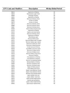
2 What is the differential diagnosis? Differential Diagnosis Obstructing renal or ureteral stone Hydronephrosis (ureteropelvic junction obstruction, stricture, ureteral/ renal malignancy) Bacterial cystitis or pyelonephritis Acute abdomen (bowel, biliary, pancreas, or aortic abdominal aneurysm) Radicular pain (L1 herpes zoster, sciatica) Depending on the patient gender, primary gonadal pathology Women: ectopic pregnancy, ovarian torsion Men: testicular torsion, orchitis What Are Some Common Types Of Kidney Stones ? Calcium oxalate Calcium phosphate concretion (called a Randall s plaque- highlighted by the arrows below), erodes through the urothelium and is a nidus for CaOx deposition.
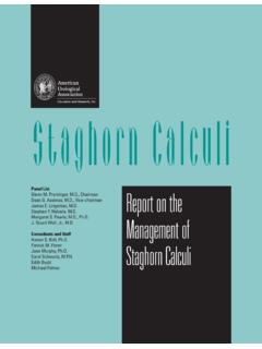
3 Risk factors: Dehydration, hypercalciuria, hyperoxaluria, hypernatrituria, hyperuricosuria. Urinary citrate is an important inhibitor of CaOx deposition. Uric Acid Stones Persistently acidic urine Persistent metabolic acidosis (eg renal tubular acidosis) Hyperuricosuria due to a variety of causes Lymphoma/ leukemia treated with chemotherapy Hyperuricemia (gout) Parallelogram shape Struvite Stones Also called magnesium ammonium phosphate stone Caused by UTIs with urease-producing organisms Commonly Proteus E. Coli is not urease-producing Urea NH4 + OH- (raises urine pH) Can form staghorn calculi which occupy the calyceal spaces/ internal renal volumetric capacity Cystine Stones Amino acid of cysteineS-S -cysteine One of the 4 dibasic amino acids including ornithine, lysine, and arginine (COLA) Cystine Stones produced in patients homozygous for recessive cystine transport gene Forms in acidic urine Hexagonal shape HOW WOULD YOU DIAGNOSE A Kidney STONE?
4 Diagnosis of a Kidney Stone Gold standard is a CT of the abdomen and pelvis without IV contrast Ultrasound is not sensitive for ureteral calculi, but is the test of choice in pregnant women A plain abdominal radiograph (KUB) can diagnose 75-90% of Stones Uric acid Stones are radiolucent and cannot be seen on KUB HOW ARE Stones MANAGED AND WHEN ARE THEY AN EMERGENCY? Situations where Stones Require Urgent Intervention Obstructed upper tract with infection (fever, elevated WBC, signs of infection on urine analysis and microscopy) Impending renal deterioration (as in a solitary Kidney ) Pain refractory to analgesics Intractable nausea/ vomiting Placement of a ureteral stent/ percutaneous nephrostomy tube to decompress the Kidney Does not involve breaking up the stone, as bacteria are often housed within the stone and this could worsen urosepsis Management in the acute setting WHAT SIZE Stones ARE LIKELY TO PASS AND WHAT ARE NON-SURGICAL TREATMENTS FOR Stones ?
5 Chance of Passing Ureteral Stones Stone Size (mm) # of days to pass stone (mean) % Likelihood of eventual need for intervention 2 or less 8 3 3 12 14 4-6 22 50 >6 -- 99% Medical Expulsion Therapy (MET) MET shortens the duration of stone passage and increases the likelihood of stone passage Includes alpha-blockers and calcium channel blockers in combination with NSAIDs Encourage hydration up to 2L/ day of fluid intake and ask the patient to strain their urine to catch and submit their stone for analysis WHAT ARE SURGICAL AND NON-SURGICAL OPTIONS FOR STONE INTERVENTION? Stone Intervention Options Oral Stone Dissolution Specific to uric acid Stones (5-10% of all urinary calculi), can be managed with urine alkalinization with potassium citrate extracorporeal shock wave lithotripsy (ESWL)
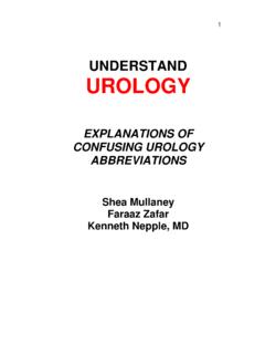
6 External shock waves are concentrated over the area of the stone Many variables at play to determine likelihood of stone clearance, but ideal for Stones <3cm and not in the lower pole Ureteroscopy and Laser lithotripsy Direct visualization and fragmentation of the stone with a laser Percutaneous Nephrolithotomy Percutaneous removal of large Stones or staghorn calculi Stone-free rates after various Urological procedures. Note: PNL- percutaneous nephrolithomy; SWL-shock wave lithotripsy . (From: Preminger et al, J Urol 2005;173:1991-2000). WHAT ARE MEASURES TAKEN TO PREVENT STONE RE-FORMATION? WHAT IS THE ROLE OF DIET?
7 Metabolic Stone Evaluation To be undertaken chiefly among patients with recurrent stone episodes and when the patient does not have an obstructing stone 24 hour urine collection for total volume, calcium, oxalate, sodium, uric acid, citrate, phosphate, magnesium, sulfate, & creatinine Serum calcium, phosphorous, uric acid, HC03, BUN, creatinine, albumin, alkaline phosphate, intact PTH (optional), 1, 25-di-OH-vitamin D2 (optional) Stone composition analysis General Dietary Guidelines Increase fluid intake & low salt diet reduces the likelihood of stone supersaturation Moderate animal protein regulates uric acid Moderate calcium a certain amount of calcium is needed in the diet to bind oxalate and prevent hyperoxaluria Increased dietary citrate- found in lemons and oranges; a major buffer for urinary pH Summary Urinary calculi typically present with renal colic and hematuria.
8 A non-contrast CT scan of the abdomen and pelvis is the best initial diagnostic test. Clinicians must assess the need for urgent intervention and the likelihood of stone passage. Metabolic risk of stone recurrences should be addressed most commonly in repeat stone formers. References Preminger GM, Assimos DG, Lingeman JE, Nakada SY, Pearle MS, Wolf JS Jr : Report on the Management of Staghorn Calculi. accessed May 7, 2011 Preminger GM, Tiselius H-G, Assimos DG, et al: 2007 Guideline for the management of ureteral calculi. accessed May 7, 2011 Preminger GM, Ferrandino MN, Haleblian GE, et al: Urology Core Curriculum: Surgical Stone Management.
9 Accessed May 7, 2011 Matlaga BR, Coe FL, Evan AP, Lingeman JE: The role of Randall's plaques in the pathogenesis of calcium Stones . J Urol 2007; 177: 31-8. Miller OF, Kane CJ: Time to stone passage for observed ureteral calculi: a guide for patient education. J Urol 1999; 162: 688-91. Preminger GM, Assimos DG, Lingeman JE, et al: Chapter 1: AUA Guideline on management of staghorn calculi: diagnosis and treatment recommendations. J Urol 2005; 173: 1991-2000. Segura JW, Preminger GM, Assimos DG et al: Ureteral Stones clinical guidelines panel summary report on the management of ureteral calculi. J Urol 1997; 158: 1915.
10 P a k C Y, Resnick MI: Medical therapy and new approaches to management of urolithiasis. Urol Cl N Am 2000; 27: 243-53.