Transcription of Klebsiella肺炎4例の画像的検討 - jrs.or.jp
1 530 40 6 2002 .. klebsiella 4 . 1 2 1 1 1 . 1 1 1 3 1 . 1 1 1 4 1 . klebsiella pneumoniae klebsiella 3 . klebsiella 1 computed tomography CT . 4 4 Ampicillin . 1 3 2 .. 1 3 . 1 .. klebsiella pneumonia Opportunistic infection Lobar pneumonia Bronchopneumonia . Aspiration pneumonia Lung abscess .. klebsiella 1 5 20 ! 60 . 12 9 28 . 1 . X . 165 cm . 2 54 kg 30 ! 124 ! Friedlander turgor . com- . puted tomography CT 3 mm3 88 94. 17,000! klebsiella pneumoniae mm! hr CRP mg! dl BUN mg! dl Cr mg! 2000 dl room air PaO2 4 . 4 mmHg PaCO2 mmHg 9 28 . klebsiella klebsiella 3 X air klebsiella 1 CT bronchogram Fig.
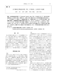
2 1a . CT . Fig. 1b .. Cefoperazone! sulbactam 2 g! . day Clindamycin phosphate 1,200 mg! day . 1 89 Methiciline-resistant staphylo- coccus aureus MRSA 4 .. 390 8621 3 1 1. 1 .. 1 . 2 klebsiella .. 3 . 2 54 . 4 .. 13 9 19 46 . 531. b Fig. 1b Case 1. CT of the right lower lobe showing a a dense, confluent infiltrate, involving almost the entire Fig. 1a Case 1. Posteroanterior PA chest radiograph lobe. Emphysematous bullae are also present in the in Sep. 2000 demonstrating a sign of open bronchus, middle lobe. with signs of air-bronchogram indicating alveolar dis- ease in the right lower lung field.
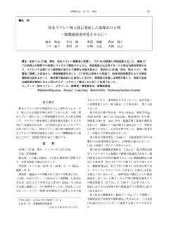
3 B a Fig. 2b Case 2. A lung field CT image showing patchy and irregular opacity in the right upper lobe. These Fig. 2a Case 2. PA chest radiograph in Jul. 2001. acute infiltrates indicate bronchopneumonia in S2. shows patchy infiltrations in the right upper lung field and in both lower lung fields.. 13 7 24 mm3 . 12,700! Clarithromycin 81 . 92 mm! hr CRP mg! dl HbA. 27 1c room 148 cm 41 kg 27 ! air PaO2 mmHg PaCO2 mmHg . 118 ! 142 mg! dl 7 26 . 532 40 6 2002 . b a Fig. 3b Case 3. A lung field CT image showing acute bronchopneumonia with peribronchial ground glass Fig.
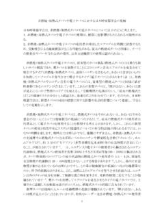
4 3a Case 3. PA chest radiograph in Aug. 2001. opacity in the left upper lobe. showing diffuse infiltration in the left lung field. X Fig. 3b Ceftazidime 2 g! day Clindamy- Fig. 2a . CT cin phosphate 1,200 mg! day 2 . Fig. 2 . b . Ampicillin! sulbactam 6 g! day 3 Citrobac- 2 ter freundii 2 . klebsiella . 3 Enterobacter cloacae 1 . 4 70 . klebsiella . 3 74 65 . 20 ! 45 1 2 . 72 . 13 5 .. 13 7 17 . 13 8 16 X .. 144 cm 38 kg 156 cm 48 kg 24 ! . 28 ! 102 ! 98 ! valve click .. mm3 86 62. 33,500! mm3 . 9,730! mm! hr CRP mg! dl 76 88 mm! hr CRP mg! dl GOT. O2 3 l PaO2 mmHg PaCO2 57 U!
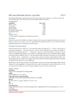
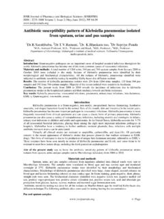
5 L GPT 46 U! L ZTT kunkel TTT kunkel mmHg 8 16 X room air PaO2 Fig. 3a . 8 21 mmHg PaCO2 mmHg 7 17 . high-resolution CT HRCT X 2 . Fig. 4a . CT . 533. a b Fig. 4a Case 4. PA chest radiograph in July 2001. showing a consolidation in the right lung. Two cavitations containing debris appear to be pre- sent in the consolidation. Fig. 4b Case 4. A lung field CT image in which dense consolidation can be identified in the mid- dle lobe. Fig. 4c Case 4. A lung CT at a different window setting, revealing a central low-density area con- taining air, which is an abscess.
6 A small right pleural effusion is also present. c Fig. 4b .. 5 . Fig. 4 c . 2 10 6 . 7 . klebsiella . klebsiella . Ciprofloxacin 600 mg! day .. 4 . klebsiella pneumoniae 1 3 . 534 40 6 2002 . klebsiella CT 1 .. 2 . 1 . 3 3 1989 ; 572 574. X 2 Macfarlane JT : Treatment of lower respiratory in- HRCT fections. Lancet 1987 ; 8573 : 1446 1449. 3 Moon WK, Im JG, Yeon KM, et al : Complications of 4 klebsiella . klebsiella pneumonia : CT evaluation. J Comput As- debris . sist Tomogr 1995 ; 19 : 176 181. 8 9 . 4 . CT .. 2000 ; 20 21. 5 6 . 10 1998 ; 361. 3 . 11 . klebsiella 6.
7 2001 ; 49 : 731 737.. 7 .. prospective study 1996 ; 34 : 759 764. 8 1 . 1995 ; 197. 1 12 13 9 . Kleb- extended-spectr- siella 2001 ; 39 : 419 423. um 14 . beta-lactamase : ESBL klebsiella 10 Carpenter JL. klebsiella pulmonary infections : oc- currence at one medical center and review. Rev In- Metallo-beta-lactamase . fect Dis 1990 ; 12 : 672 682. klebsiella 15 . 11 klebsiella pul- 4 . mononiae . 1 2001 ; 39 : Carbapenem Newquinolone 405 409. 16 12 MRSA .. Medical View 1997 ; 309 311. 13 . 1998 ; 87 : 243 248.. 14 . 17 I . ESBLs 2001 ; 59 : 694 700. II III . 15.
8 4 klebsiella 2001 ; 59 : 701 706. CT 16 Gilbert DN, Moellering RC, Sande MA : The sanford 3 guide to antimicrobial therapy 2001 31 th edition .. 2 Antimicrobial Therapy, Inc., Main Street Hyde Park, VT, 2001 ; 54. 17 2001 ;. 20 : 692 699. 535. Abstract Four Cases of klebsiella pneumonia Akihiro Tsukadaira1 , Yoshio Okubo2 , Takashi Kobayashi1 , Toshihide Wakamatsu1 , Mari Sasabayashi1 , Junichi Hotta1 , Kenji Tsushima1 , Shuji Takashi1 , Yoshitaka Yamazaki3 , Shinji Yamaguchi1 , Masayuki Hanaoka1 , Tomonobu Koizumi1 , Keisaku Fujimoto1 , Siro Horie4 and Keishi Kubo1.
9 1 . The First Department of Internal Medicine, Shinshu University School of Medicine, Asahi 3 1 1, Matsumoto, 390 8621. 2 . Department of Internal Medicine, Higashinagano National Hospital, Uwano 2 477, Nagano, 381 8567. 3 . Division of Endoscopy, Shinshu University Hospital, Asahi 3 1 1, Matsumoto, 390 8621. 4 . Department of Internal Medicine, Okaya Enrei Hospital, Okaya 4769,394 8588. Typical klebsiella pneumonia with mucous sputum is known as an opportunistic nosocomial infection. How- ever, computed tomographic study of limiting in klebsiella pneumonia is rare.
10 We report three types of chest com- puted tomography CT findings for klebsiella pneumonia. Case 1 shows typical lobar pneumonia Friedlander pneumonia ,Cases 2 and 3 show acute bronchopneumonia with subclinical aspiration, and Case 4, chronic Kleb- siella pneumonia with typical cavitary lung abscesses. Of these four cases of klebsiella pneumonia, three devel- oped in the right lung, as determined radiologically, but esophagogastroduodenoscopy indicated that the lesions of Case 3 had developed in the left lingula and upper lobe.