Transcription of Lab #11: Respiratory Physiology
1 Lab #11: Respiratory Physiology Background The Respiratory system enables the exchange of O2 and CO2 between the cells and the atmosphere, thus enabling the intake of O2 into the body for aerobic respiration and the release of CO2 for regulation of body fluid pH. In this exercise, we will examine ventilation of the lungs to enable the exchange of air between the alveoli and the atmosphere, and also explore the rate of CO2 release from the lungs as a mechanism for controlling pH. Fig Relaxation of the inspiratory muscles drives tidal expiration (left), whereas forced expirations are driven by contraction of the expiratory muscles (right). Mechanics of Lung Ventilation. From L. Sherwood, Fundamentals of Human Physiology . Brooks Cole. Air flow into and out of the lungs is driven by The net result is an increase in the volume of the pressure differences between the atmospheric air thoracic cavity.
2 Since the lungs are adhered to and air in the lungs. When atmospheric pressure the inner walls of the thoracic cavity, the lungs exceeds intrapulmonary pressure, air flows into also expand. This decreases intrapulmonary the lungs, and when intrapulmonary pressure pressure below atmospheric pressure, and air exceeds atmospheric pressure, air flows out of flows into the lungs along the pressure gradient. the lungs. Changes in intrapulmonary pressure Tidal expiration is driven by the relaxation are driven by changing the volume of the of the muscles used to drive tidal inspiration lungs per Boyle's Law, the pressure exerted by (Fig ). As the diaphragm moves upward a given amount of gas at a constant temperature and backward and the ribs move downward and is inversely proportional to the volume of that inward the overall volume of the thoracic cavity gas. Thus increasing the volume of the lungs is reduced.
3 This compression of the thoracic will decrease intrapulmonary pressure, whereas cavity, in turn, elevates intrapulmonary pressure decreasing lung volume will increase above atmospheric pressure, and air flows out of intrapulmonary pressure. the lungs. During tidal ventilation, air is inspired by Additional amounts of air can be inspired or contracting certain muscles in the walls and expired from the lungs with the contraction of floor of the thoracic cavity (Fig ). As the additional muscles. Maximal inspirations are diaphragm contracts, it pulls downward and driven with contraction of muscles associated forward, whereas when the external intercostals with the sternum and clavicle in addition to a contract, they lift the ribs upward and laterally. full contraction of the diaphragm and external intercostals. Air can be forcibly expired from the lungs with contraction from the internal intercostals and a number of abdominal muscles (Fig ).
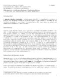
4 Lung Volumes and Capacities. The amount of air contained in the lungs during ventilation can change considerably depending Fig Muscle contractions that drive tidal on what muscles are driving air flow and how inspiration. From L. Sherwood, Fundamentals of forcefully they contract. The different amounts Human Physiology . Brooks Cole of air drawn into or out of the lungs by Lab #11: Respiration Residual Volume (RV). The residual volume is the amount of air that remains in the lungs following a maximal 6000. IRV expiration, and can only be forced out of IC the lungs by collapsing the lungs. The Volume (ml). VT. VC RV for men is approximately 1200 ml TLC and approximately 900 ml in women. ERV. FRC. RV There are also four lung capacities, each of 0 which is the sum of two or more primary lung Primary Lung Volumes Lung Capacities volumes. Fig. Illustration of a spirometer recording Total Lung Capacity (TLC).
5 The total depicting measurements of the primary lung volumes (left) and the lung capacities (right). lung capacity is the maximum amount of air that can be held within the lungs at one time, and is the volume of air in the contracting different groups of muscles are lungs following a maximal inspiration. called primary lung volumes. Different TLC is the sum of all four primary combinations of the primary lung volumes, in volumes (TLC = IRV + VT + ERV. turn provide us with lung capacities, which +RV). define either how much air is present in the Vital Capacity (VC). The vital capacity lungs or how much air can be moved by the is the maximum amount of air that can lungs under specific situations. be exchanged between the lungs and There are four primary lung volumes (Fig atmosphere in a single breath, and is the ), defined as follow. volume of air that can be forcibly expired from the lungs following a Tidal volume (VT).
6 The tidal volume is maximal expiration. The vital capacity the amount of air inspired (or expired). is the sum of the three primary volumes during normal tidal breathing. At rest, that can be directly exchanged with the tidal volume in healthy adult men is atmosphere (VC = IRV + VT + ERV). approximately 500 ml, and about 400 ml Inspiratory Capcity (IC). The in women. Tidal volume increases with inspiratory capacity is the maximum activity to accommodate increased need amount of air that can be inspired for gas exchange. following a normal tidal expiration. It is Inspiratory Reserve Volume (IRV). The the sum of the inspiratory reserve inspiratory reserve volume is the volume volume and the tidal volume (IC = IRV. of air that can be maximally inspired + VT). above the volume inspired tidally. Functional Residual Capacity (FRC). Average IRV measurements at rest for The functional residual capacity is the men and women are approximately 3100.
7 Volume of air that remains in the lung ml and 2400 ml, respectively. IRV. following a normal tidal expiration. It is decreases with exercise. the sum of the expiratory reserve Expiratory Reserve Volume (ERV). The volume and the residual volume (FRC =. expiratory reserve volume is the ERV + RV). maximum volume of air that can forcibly expired beyond a normal tidal expiration. Average ERV at rest is Measurements of Tidal Ventilation. approximately 1200 ml for men and 900. ml for women at rest. Like IRV, the The amount of air exchanged between the lungs ERV decreases when exercising. and the atmosphere during tidal breathing is Lab #11: Respiration 5000 where VDS is the volume of the dead space 4000 (estimated to be 1/3 of the resting tidal volume). Volume (ml). 3000. 2000 Air Flow Measurements. 1000. Efficient air exchange between the lungs and the 0. atmosphere requires a) that the lungs be able to change volume effectively and b) air can pass 0 1 2 3 through the Respiratory passageways with FEV1 = (5000 ml -1000 ml) / 5000ml = 4000 ml / 5000 ml Time (sec) relative ease.
8 The ability of a person's lungs to = 80% change in volume is reflected in their vital capacity measurement individuals with larger Fig Illustration of a spirometer recording vital capacities can change the volume of their measuring forced expiratory volume. lungs more that can those with smaller vital determined influenced how much air is capacities. The ability of air to flow through the exchanged in each breath (the tidal volume) and Respiratory passages, in contrast is reflected in a how frequently breaths are take (the breathing measurement called the forced expiratory rate). Measurements of tidal ventilation, volume (FEVt), which is the percentage of the therefore, must take both of these factors into vital capacity that, after a maximal inspiration, account. One simple measurement of tidal can be forcibly expired in t seconds (Fig ). ventilation is the minute volume (VM), which is FEVt can be calculated as the ratio of air the volume of air inspired through tidal forcibly expired in a designated time interval breathing during in a one minute period of time.
9 (Vt, not to be confused with tidal volume, VT). Minute volume is simply the product of the tidal divided by the vital capacity (VC) and converted volume (VT) and the breathing rate (f) into a percentage. VM = f VT Vt FEVt = 100%. VC. The minute volume, however, overestimates the amount of air that is available for gas exchange. A young adult can typically expire roughly 80%. This is because not all of the air flowing into the of their vital capacity within one second, 94%. lungs during inspiration flows into the alveoli; within two seconds, and 97% within three some of this air accommodates the increased seconds. volume of the Respiratory passages during Vital capacity and FEVt measurements can inspiration, and since these passages are not be used to diagnose various types of air flow designed for gas exchange with the blood, they disorders. Abnormally low FEVt measurements are considered to be physiological dead space.
10 (less than 90% the value predicted for an A better measurement for the amount of air individual based on their age) may be indicative flowing over the Respiratory surfaces during tidal of an obstructive disorder. In an obstructive ventilation is alveolar ventilation (VA), disorder, air flow through the Respiratory sometimes called the minute alveolar volume, passages is impeded by a narrowing of those which is the amount of air entering the alveoli in passages, thus increasing resistance to air flow. a one minute period. Alveolar ventilation is Bronchiolar secretions and constriction of air calculated using a similar equation to that of the passageways (See Fig ) are common causes minute volume except that it corrects for the of obstructive disorders. On the other hand, volume of the dead space: abnormally low vital capacity (less than 80% the predicted value based on age, sex, and body VA = f (VT - VDS) size) may be indicative of a restrictive disorder.