Transcription of LECTURE 4 - MICROSCOPIC STUDY OF MINERALS UNDER THE …
1 LECTURE series: SGL 201 Principles of Mineralogy. 63 LECTURE 4 INTRODUCTION TO THE MICROSCOPIC STUDY OF MINERALS LECTURE OUTLINE Welcome to the fourth LECTURE of this unit. You should now be familiar with the concepts of optical mineralogy covered in LECTURE 3. In this LECTURE , we are going to learn firstly about the important components of the petrographic microscope itself. Technologically, microscopes vary in their design, not only in their appearance but also in the positioning and operation of the various essential components. These components are present in all microscopes and will be described briefly in this LECTURE . Although dual purpose microscopes incorporating both transmitted and reflected light options are now available in the market, its is more convenient for the purpose of this unit and for your ease of understanding, to describe the two techniques separately.
2 Secondly, in learning about the MICROSCOPIC STUDY of MINERALS , we shall give a systematic description of the characteristic optical properties observed UNDER plane- and crossed-polarized light. Finally we shall review the general procedures used in the making of thin- and polished sections. At the end of this LECTURE , you should be able to: Describe and illustrate the use of all the major components of the transmitted light polarizing microscope Give a systematic description of MINERALS in thin sections using transmitted light. Explain the optical properties observed UNDER plane-polarized light and crossed polars. Give examples of MINERALS with distinctive optical properties Describe the use of the Reflecting Microscope.
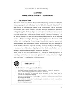
3 Describe the general procedures of making thin- and polished-sections. OBJECTIVES LECTURE series: SGL 201 Principles of Mineralogy. THE TRANSMITTED-LIGHT MICROSCOPE The polarizing transmitted-light microscope, commonly known as petrographic microscope, consists essentially of a light source, a sub-stage condenser and a stage to hold the specimen, an objective, and an eyepiece (Figure ). In addition to these, there is a device for producing polarized light, termed the polarizer, a graduated rotating stage as opposed to a fixed stage, and a second polarizing device termed the analyser. A brief description of these major components is discussed here below. The light source In transmitted-light studies a lamp is commonly built into the microscope base (Figure ).
4 The typical bulb used has a tungsten filament, which gives the field view a yellowish tint. A blue filter can be inserted immediately above the light source to change the light colour to that of daylight. Figure The transmitted-light polarizing microscope LECTURE series: SGL 201 Principles of Mineralogy. 65In older microscopes the light source is quite separate from the microscope and is usually contained in a hooded metal box to which can be added a blue glass screen for daylight colored light. A small movable circular mirror, one side of which is flat and the other concave, is attached to the base of the microscope barrel. The mirror is used to direct the light through the rock thin section on the microscope stage, and the flat side of the mirror should be used when a condenser (described below) is present.
5 The polariser The assumption is that light consists of electromagnetic vibrations. These vibrations move outwards in every direction from a point source of white light, such as a microscope bulb. A polarizing film (the polariser) is held within a lens system located below the stage of the microscope and this is usually inserted into the path. On passing through the polariser, the light is polarized and now vibrates in a single plane. This is called plane-polarized light (PPL). In most UK microscopes the polariser is oriented to give an E-W vibrating incident light. Substage diaphragms One or two diaphragms may be located below the stage.
6 The field diaphragm, often omitted on simple student microscopes, is used to reduce the area of light entering the thin section, and should be in focus at the same position as the thin section: it should be opened until it just disappears from view. The aperture diaphragm is closed to increase resolution, it can be seen when the Bertrand s lens is inserted. The condenser or convergent lens A small circular lens (the condenser) is attached to a swivel bar, so that it can be inserted into the optical train when required. It serves to direct a cone of light on the thin section and give optimum resolution for the objectives (described below) used. The entire lens system below the microscope stage, including polarizer, aperture diaphragm and condenser, can often be racked upwards or downward in order to optimize the quality of illumination.
7 LECTURE series: SGL 201 Principles of Mineralogy. Stage The microscope stage is flat and can be rotated. It is marked in degree units, and a side vernier enables angles of rotation to be accurately measured. The stage can usually be locked in place at any position. The rock thin section is attached to the centre of the stage by metal spring clips. Objectives Objectives are magnifying lenses with the power of magnification inscribed on each lens ( x5, x30). An objective of very high power ( x100) usually requires immersion oil between the objective lens and the thin section. Eyepiece The eyepiece (or ocular) contains crosswires, which can be independently focused by rotating its uppermost lens.
8 Eyepieces of different magnification are available. Monocular heads are standard on student microscopes. Binocular heads may be used and, if correctly adjusted, reduce eye fatigue. The analyzer The analyzer is similar to the polariser; it is also made of polarizing film but oriented in a N-S direction, at right angles to the polariser. When the analyzer is inserted into the optical train, it receives light vibrating in an E-W direction from the polariser and cannot transmit this; thus the field of view is dark and the microscope is said to have crossed polars (CP, XPOLS or XP). With the analyzer out, the polariser only is in position; plane polarized light is being used and the field of view appears bright.
9 The Bertrand lens This lens is used to examine interference figures (see section ). When it is inserted into the upper microscope tube an interference figure can be produced which fills the field of view, provided that the convergent lens (condenser) is also inserted into the optical path train. LECTURE series: SGL 201 Principles of Mineralogy. The accessory slot Below the analyzer is a slot into which accessory plates, a quartz wedge, or first order red (gypsum plate), can be inserted. The slot is oriented so that accessory plates are inserted at 45o to the cross wires. In some microscopes the slot may be rotatable. Focusing The microscope is focused either by moving the microscope stage up or down (newer models) or by moving the upper microscope tube up or down (older models).
10 Both coarse and fine adjusting knobs are present. SYSTEMATIC DESCRIPTION OF MINERALS IN THIN SECTION USING TRANSMITTED LIGHT Description of the optical properties of each mineral includes those that are determined in plane-polarized light (PPL) and those that are determined with crossed polars. For most properties, a low power objective is usually used (up to x 10). Properties in plane polarized light The analyzer (see above in section ) is taken out of the optical path to give a bright image. Colour MINERALS show a wide range of colour ( the natural or body colour of a mineral), ranging from colorless MINERALS ( quartz and feldspars) to colored MINERALS ( brown biotite, yellow staurolite and green hornblende).