Transcription of Left-Sided Leg Edema Of The Elderly: A Common Variant Of ...
1 Left-Sided Leg Edema Of The elderly : A Common Variant Of The Iliac Compression Syndrome Philip D. Sloane, MD, MPH, Ruth Baldwin, PhD, Royce Montgomery, PhD, Franklin Hargett, MD, MPH, and Abraham Hartzema, PharmD, MSPH Abslrtlcl: IltIcIIgroutul: Anatomical1y, the right Common iliac artery crosses the left iliac vein and 118 accompanying lymphatics. We hypothesized that this situation could lead to a predominance of Edema , telangiectasis, and venous varicosities on the left lower ex1remities of older persons. Metbotls: To test this hypothesis, a research assislant who was blinded to the study pis examined 215 predominandy elderly reslden18 of North Carolina homes for the aged and disabled. ReSlllts: Among these subjects, percent had predominantly Left-Sided Edema , and percent had predominandy right-sided Edema (P < ). When the 88 subjects with pitting Edema greater than 3 mm were studied, percent showed a Left-Sided predominance, and percent showed a rtpt-sided predominance (P < ).
2 In contrast, no signif:lcant cWJerenc:e was found in the Iateralization ofvenous varic:osities or of telangiectasis. CtmcIfISIou: Asymmetric: Edema is Common and is usually Left-Sided in older persons. Compression of die left Common Wac vein and i18 accompanying lymphatics by the right iliac artery, rather than overt c:linic:al disease, might explain the majority of asymmetric: Edema seen in c:linic:al praetic:e. (J Am Board Fam Pract 1993; 6:1-4.) Physicians in internal medicine, general practice, family medicine, and a variety of medical and surgical subspecialties often encounter patients with peripheral Edema and other physical signs of venous disease. Examination of such patients can be extensive, because these conditions can arise from systemic or local causes. When Edema or varicosities are unilateral or asymmetric, causes local to the involved extremity are implicated. The most Common causes of unilateral or regional leg Edema are reported to be chronic venous insuffi-ciency and ,2 Often medical textbooks recommend an intense search for causes of lymphatic obstruction in such patients, such as malignancy in the inguinal nodes or deep venous Submitted, revised, 18 August 1992.
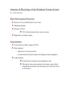
3 From the Department of Family Medicine, School of Medicine (pDS, FH); School of Phannacy (RB, AH); and Department of Anatomy, School of Medicine (RM); University of North Carolina at Chapel Hill. Address reprint requests to Philip D. Sloane, MD, MPH, Department of Family Medicine, The Uni-versity of North Carolina, Campus Box 7595, Chapel Hill, NC 27599-7595. This research was supported in part by a grant from the John Hartford Foundation. Dr. Sloane is a recipient of Academic Award No. I KOS AGOO341-01 from the National Institute on Aging. We had observed informally that venous disease was more Common on the left side, yet we found no published reports to confirm that observation. Anatomically, such an occurrence is logical, be-cause the right iliac artery crosses the venous and lymphatic drainage system returning from the left leg (Figure 1). We reasoned that, with age, any compression produced by the right iliac artery might be manifest as an increased occurrence and greater severity of venous varicosities, telangiecta-sis, and Edema on the left side when compared with the right.
4 To test our hypothesis, we conducted system-atic observations on the peripheral vascular status of 215 residents of North Carolina rest homes. These observations were performed by a blinded observer who was gathering observational data as part of a larger, unrelated study. Methods The data for this project were collected as part of a larger study conducted by the University of North Carolina Center for Health Promotion and Disease Prevention of homes for the aged (HAs), a type of domiciliary care facility. The purpose of the larger study was to improve the management of medications in HAs through educational inter- Edema of the elderly 1 J RIGHT Common ---,.t.~i:if'- ILIAC ARTERY Common ILIAC LYMPH NODES LEFT Common ~~ft-ILIAC VEIN Figure 1. Anatomic relation of the Common Uiac arteries and veins and their associated lymphatic vessels. CrossinR of the left Uiac vein and i1s accompanying lymphatic channels by the right Uiac artery could lead to increased pressures in the venous and interstitial systems of the left leg reladve to the right.
5 Vention. 1be study sampling frame consisted of the 194 HAs with more than 20 beds located in North Carolina counties with populations of at least 50,000. A two-stage stratified cluster sampling method was used to obtain a sample of 35 homes. After homes were selected, between 16 and 26 residents in each home were enrolled in the study. The inclusion criteria for residents were use of two or more medications, at least one of which was a psychoactive, an antihypertensive, or an anti-parkinsonian agent. A total of 826 residents were selected, of which 746 were enrolled. Of these, a subsample was randomly selected for study using direct (observational) methods to verify the indi-rect (reported) measures that constituted the pri-mary outcome variables of the study. These direct observations included evaluation of mental status, mobility, level of consciousness, and the presence or absence of signs of tardive dyskinesia. The data reported here were collected as part of these direct observations.
6 A standardized protocol was designed by the investigators for recording observational data on varicosities, Edema , and telangiectasis. Edema was defined as soft tissue swelling that pits; varicose veins were defined as visible veins greater than 5 mm observed above the ankle; telangiectasis was defined as a network of bluish, black, or red vessels, usually tortuous, observed on or below the ankle. Before going into the field, the research assistant performing direct observations was trained by the primary author (PDS) in use of the protocol; training involved explanation of the protocol and joint physical as-sessments in a local nursing home. 1be data col-lector was not apprised of the study's goals or hypotheses. Data from the precoded assessment forms were entered into a computerized data-base and analyzed using the Statistical Analysis System (SAS).6 To test for significant differences between groups, the McNemar test for related samples was used.
7 Results There were 215 subjects examined. Their mean age was years (range, 26 to 98 years); 75 percent were 61 years of age or older. The major-ity ( percent) were women. Racially, per-cent were white, percent were African-American, and percent were classified as other. Among the Common diagnoses were dementia ( percent), mental retardation ( percent), psychoses ( percent), hypertension ( per-cent), cardiac diagnoses ( percent), and arthri-tis ( percent). Most subjects were able to walk independently ( percent) or with assistance ( percent); percent were chair-bound or bed-bound patients. In this largely geriatric population, Edema in which 3 mm or greater of pitting could be pro-duced was present in percent of subjects table 1. Frequency and Laterality of Edema as Percentage of Subjects Observed (n = 213). Clinical Observation Pitting Edema ;;. 3 mm Ankle (n = 213) Pretibial (n = 212) Subjective comparison of leg Edema (n = 209) Laterality of Edema among subjects with.
8 3 mm leg Edema (n = 88) *McNemar's paired comparison chi-square. 2 JABFP 1993 Vol. 6 No. 1 Right Leg Left Leg Right> Left Left> Right i* PValue < < < < Thble 2. Frequency and Laterality of venous Varicosities and Telangiectasias as Percentage of SubJec:15 Observed (0 = 213). Clinical Observation Right Leg Left Leg Right> Left Left> Right i PValue Visible varicose veins Below the knee (n = 212) Above the knee (n = 169) Subjective comparison of extent of venous varicosities (n = 212) Subjective comparison of extent of telangiectasias around the ankle (n. 206) Laterality of visible varicose veins among subjects with bulging varicosities (n = 32) McNemar's paired comparison chi-square. NS not significant. tested. Whether testing was performed in the pretibial region or at the ankle, Edema of the left leg was assigned to a more severe category signif-icantly more frequently than was Edema of the right leg (Table 1).
9 The observer subjectively rated percent of subjects to have either no Edema or no asymmetry, percent to have Left-Sided Edema , and percent to have right-sided Edema . When considering the 88 sub-jects with marked Edema (pitting Edema of 3 mm or greater in either the ankle or the pretibial re-gion), this laterality of Edema was even more striking: percent exhibited Left-Sided pre-dominance, percent showed right-sided pre-dominance, and percent appeared equal. Similar Left-Sided predominance was not noted among venous varicosities and telangiectasis, however (Table 2). Although visible venous vari-cosities were Common , comparison of the extent of varicosities did not reveal statistically signifi-cant differences in laterality either below the knee, above the knee, or overall. Similarly, a subjective comparison of the extent of telangiectasis around the ankle indicated no significant difference be-tween sides. Discussion Findings from this study confirm that asymmetric, predominantly Left-Sided Edema is quite com-mon among a largely elderly population living in HAs.
10 The study failed, however, to detect laterality in the extent of venous varicosities or telangiectasis in this population. Because the data collector was not aware of the study goals or hypotheses, we believe that these NS NS NS 83 NS NS statistically significant findings represent true differences. One explanation for preferential swelling of the left leg compared with the right is anatomic. The left Common iliac vein and its accompanying lym-phatic channels must pass behind the right com-mon iliac artery, whereas their counterparts on the right flow unimpeded into the inferior vena cava (Figure 1). This anatomic configuration has been implicated as a cause of major venous thrombosis, often followed by severe peripheral Edema and postphlebitic syndrome, a rare phenomenon called the iliac compression syndrome or the May-Thurner syndrome. 7,8 In such cases, which have been primarily reported in young adults, chronic pressure by the artery is believed to lead to fibrosis of the intima of the Common iliac vein, causing partial venous obstruction.